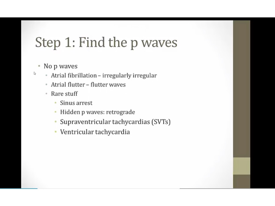
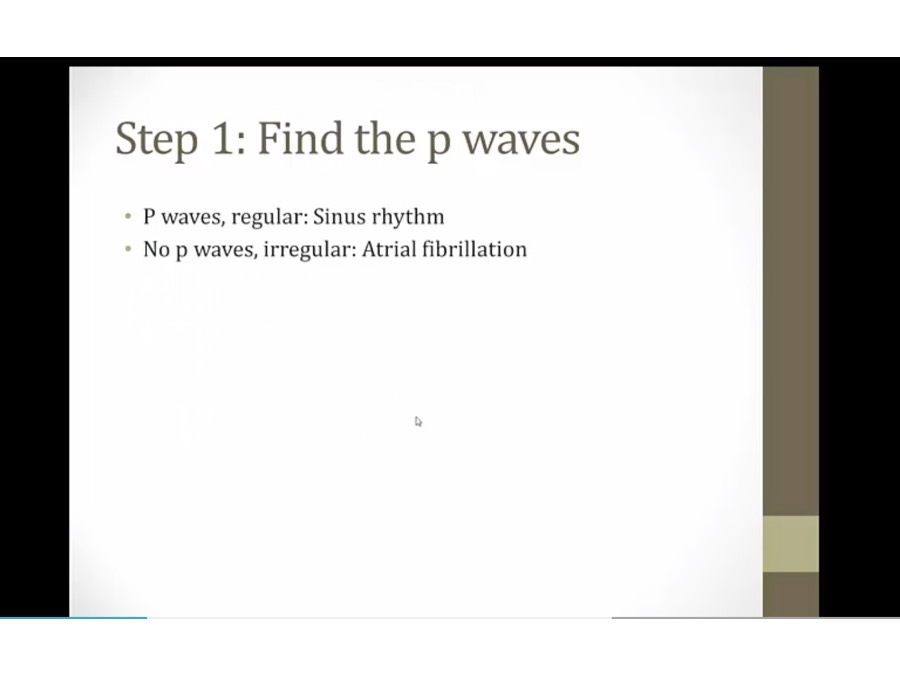
15 High Yield EKG
P
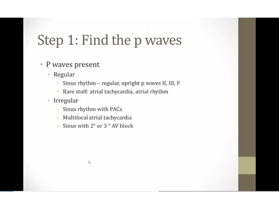
- regular: spacing between QRS same
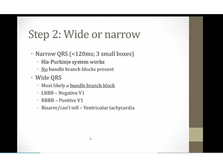
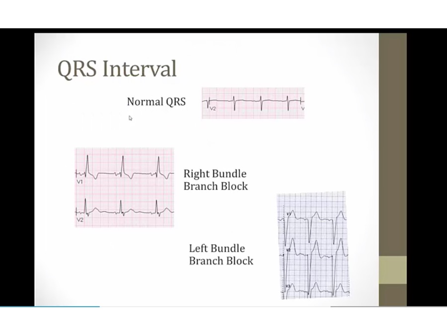
QRS
Intervals
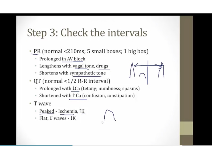
- atheletes (high vagal tones) and beta blockers prolong PR
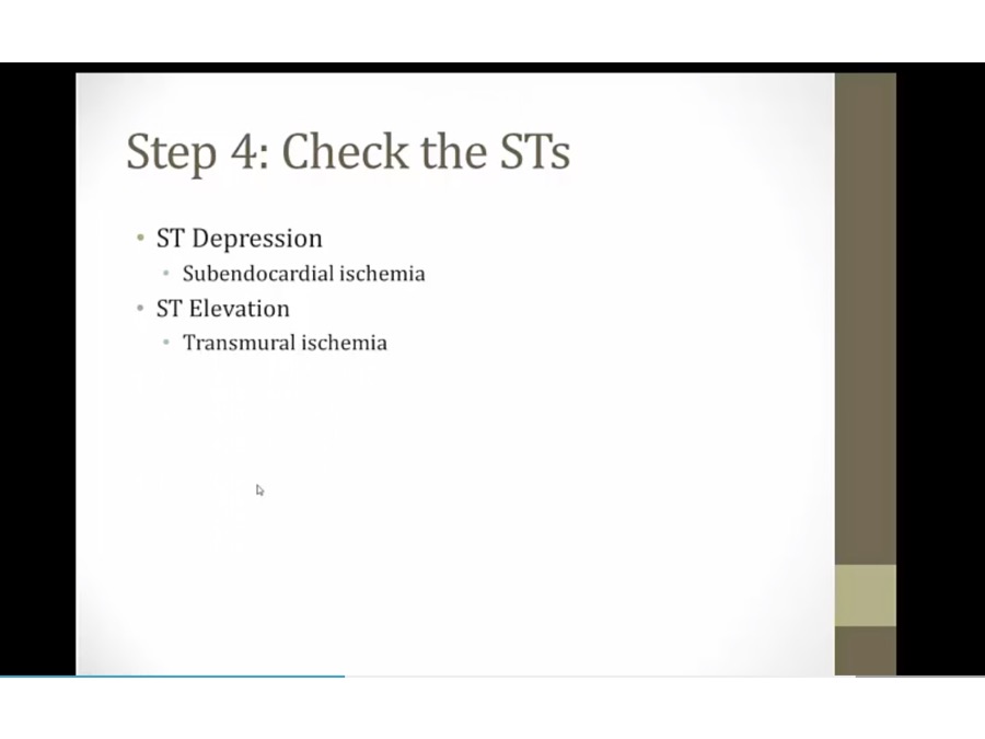
ST
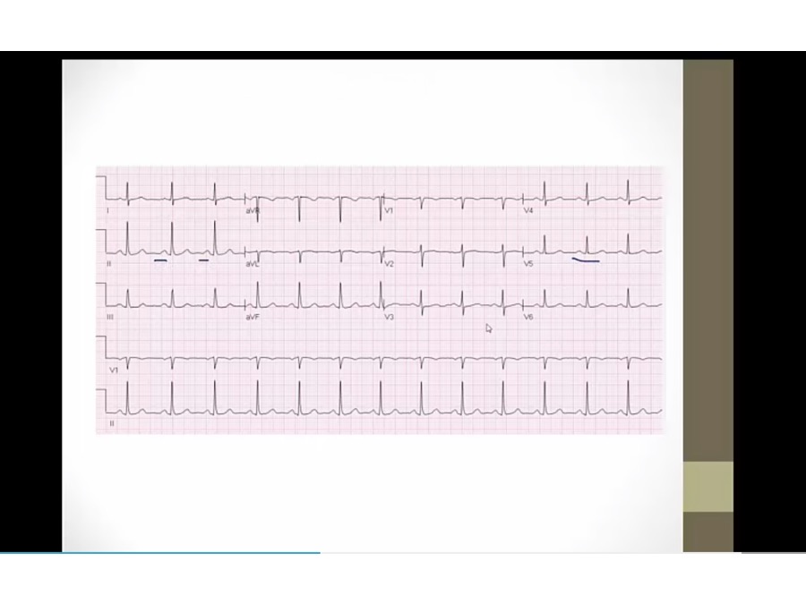
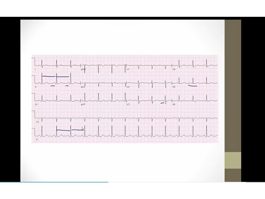
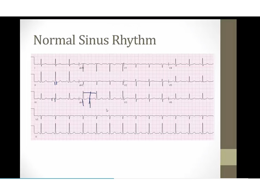
Practice 1
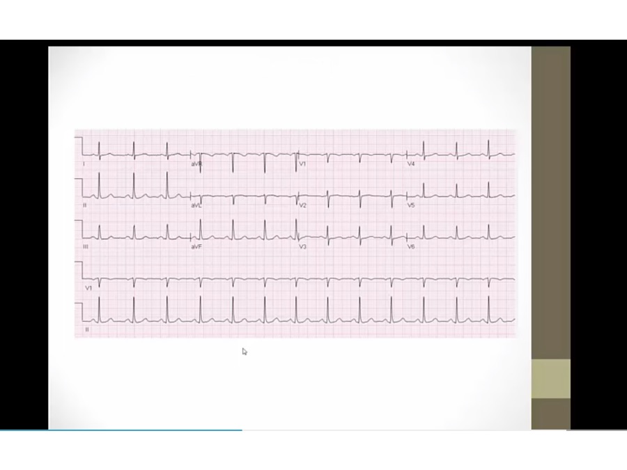
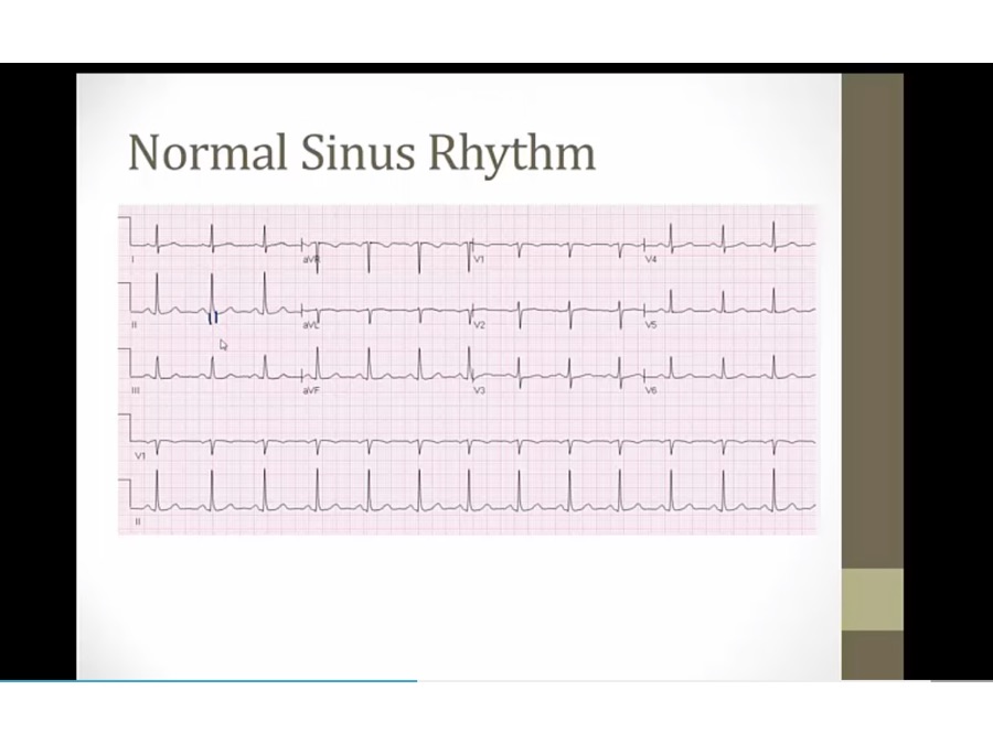
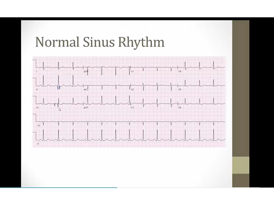
- easily identifiable P
- upright P in II, III, AVF: normal sinus P
- QRS: regular
- QRS: narrow
- PR: less than 1 block
- QT: less than half
- ST: no significant depression, elevation

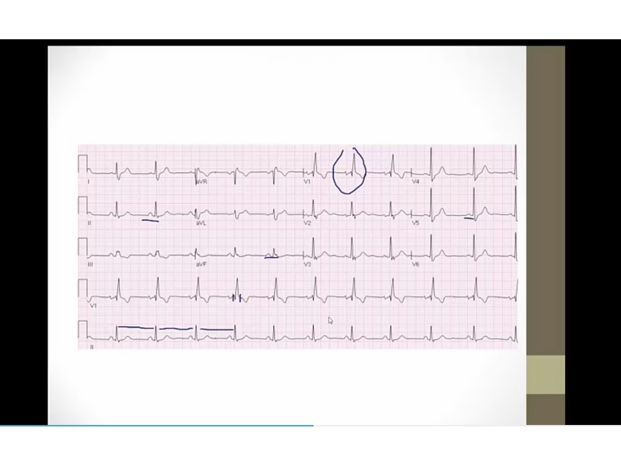
Practice 2
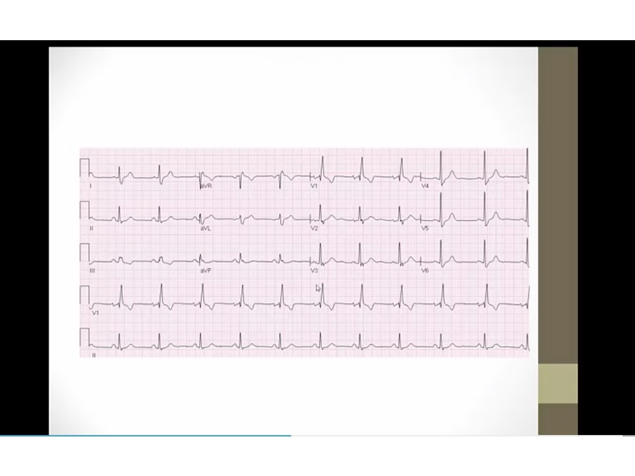
- P: upright in II, III, AVF, normal sinus P
- RR: regular QRS
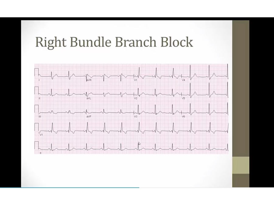
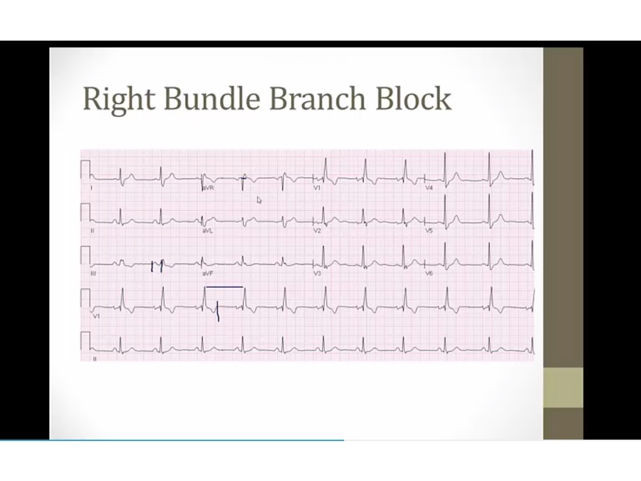
- QRS: wide in V1, upright QRS in V1 RBBB
- PR: not prolonged
- QT: less than 1/2
- ST: T inversion in V1, normal in RBBB
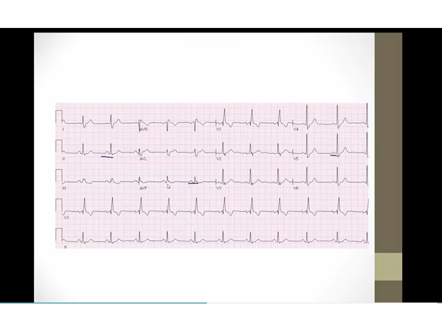


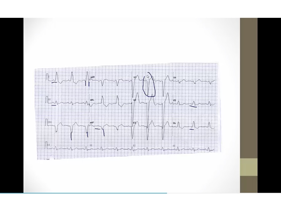
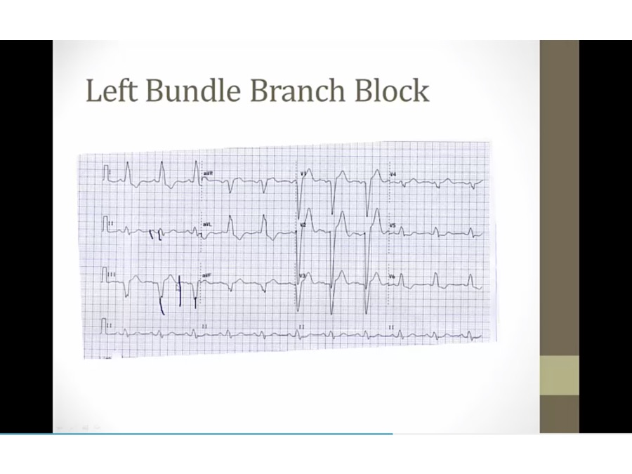
Practice 2

- P upright in II, III, AVF, sinus
- QRS wide, negative V1, LBBB
- peaked T and ST elevation in V1-3: common
- T inversion 1, L, V5, V6: normal in LBBB


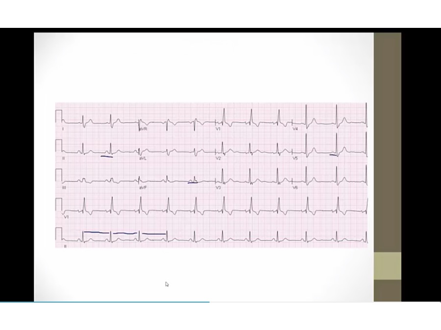
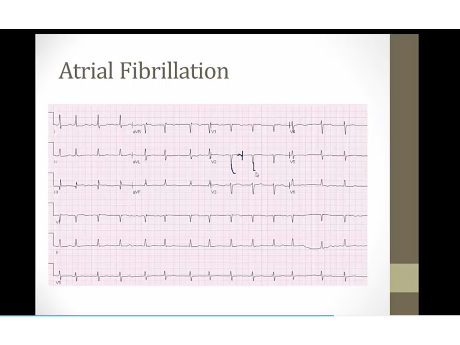
Practice 4
- no clear P waves, afib
- irregularly irregular: QRS closer in some, farther apart in others


Practice 5
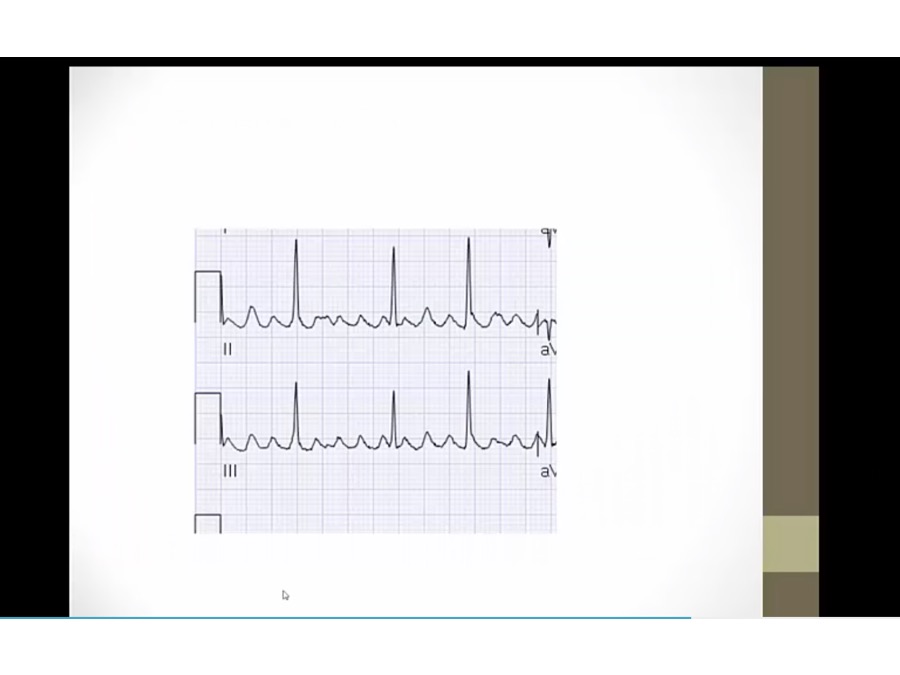
Practice 6
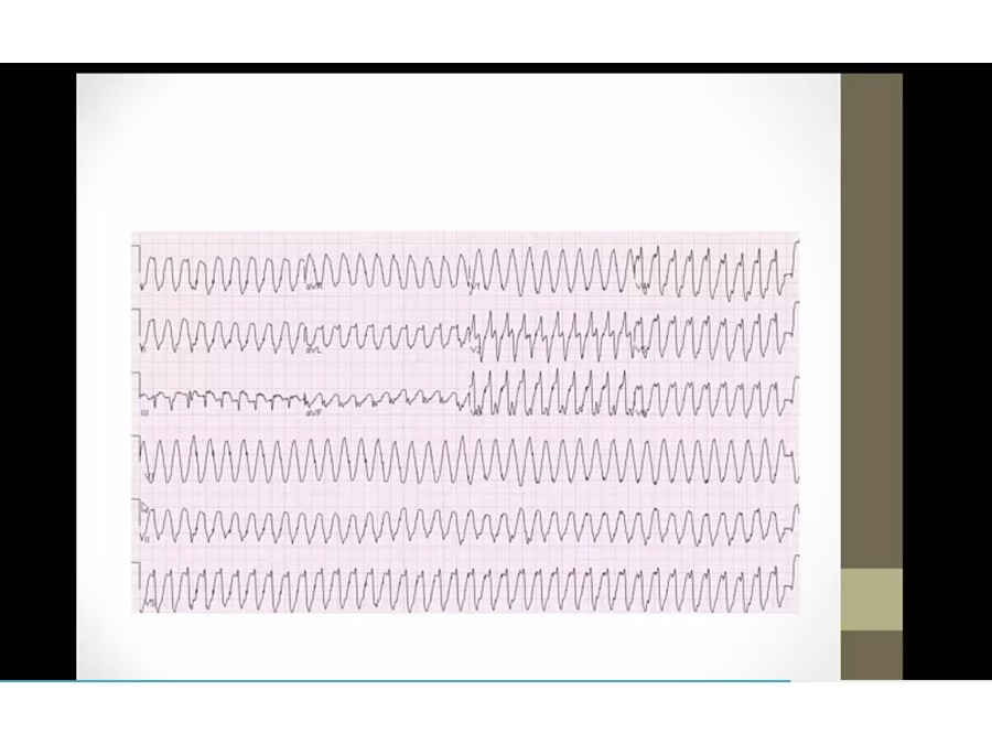
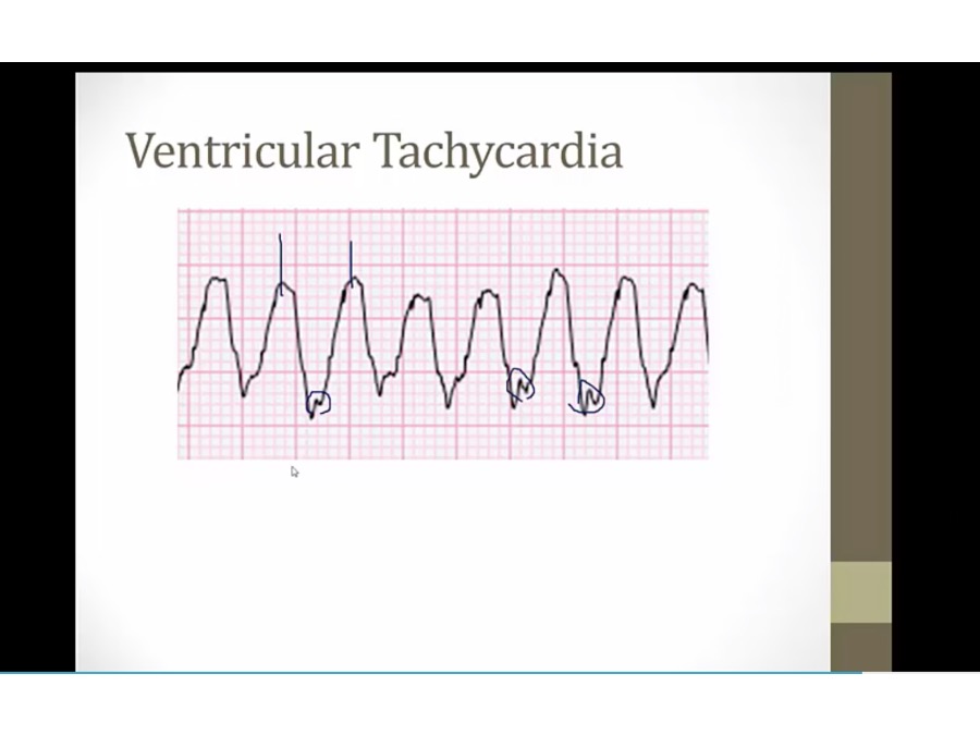
- ventricle contracts on its own
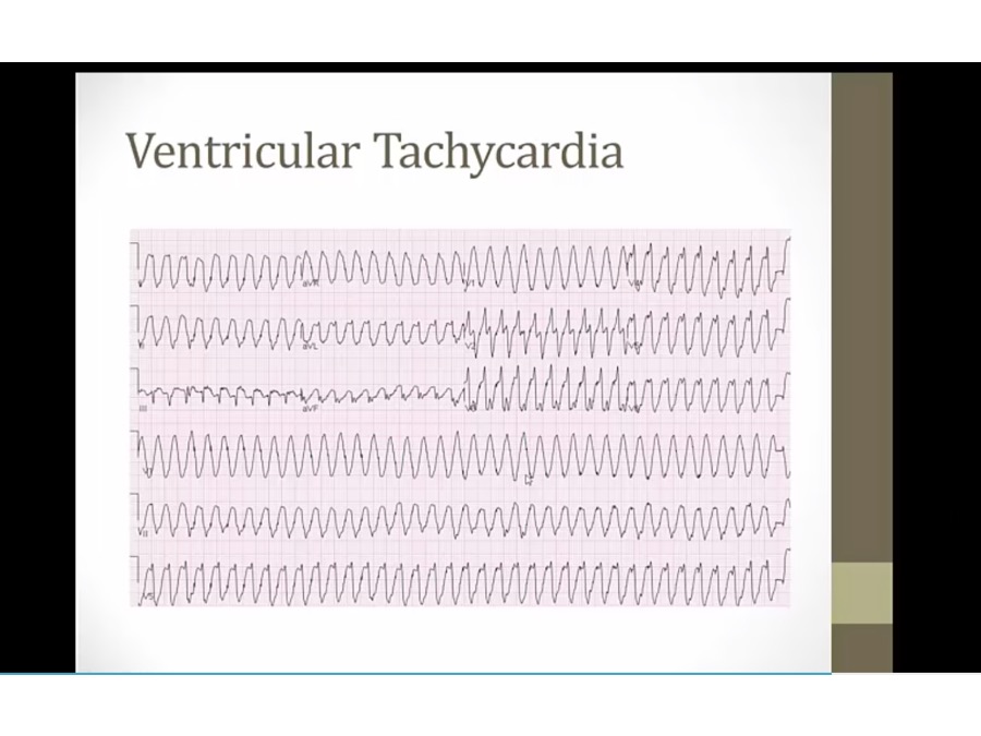
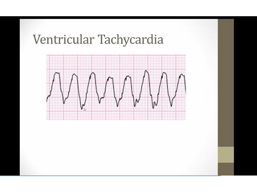
- buried P waves at different rate as QRS (AV dissociation)

VFib
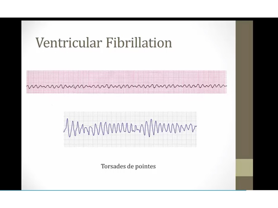
- top: VFIB, cardiac arrest
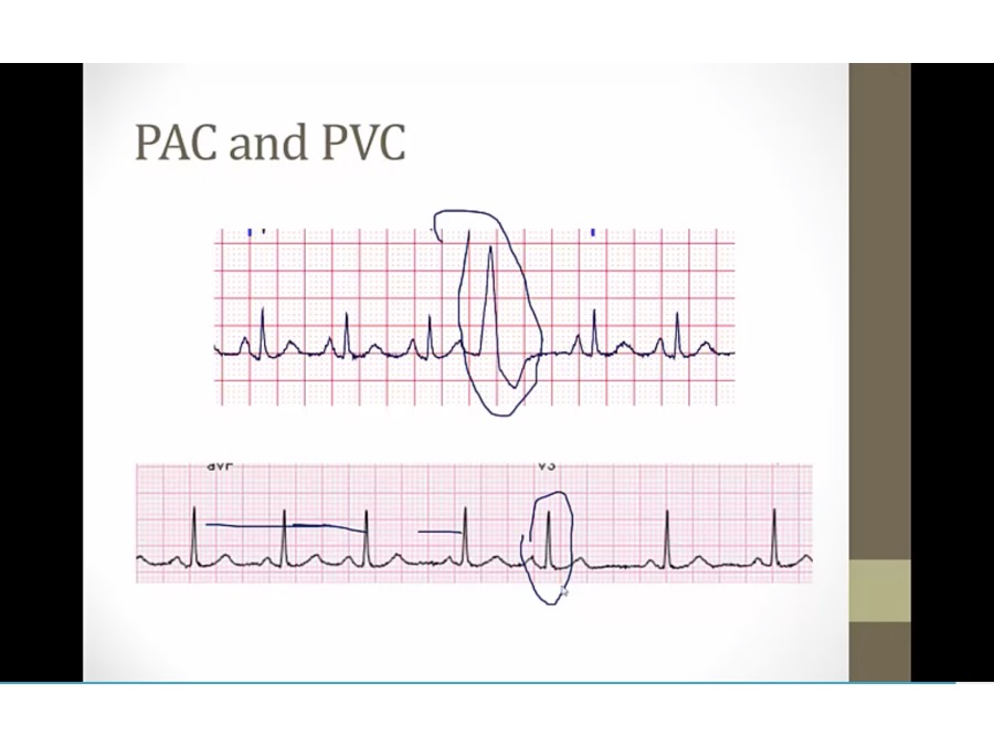
PAC, PVC
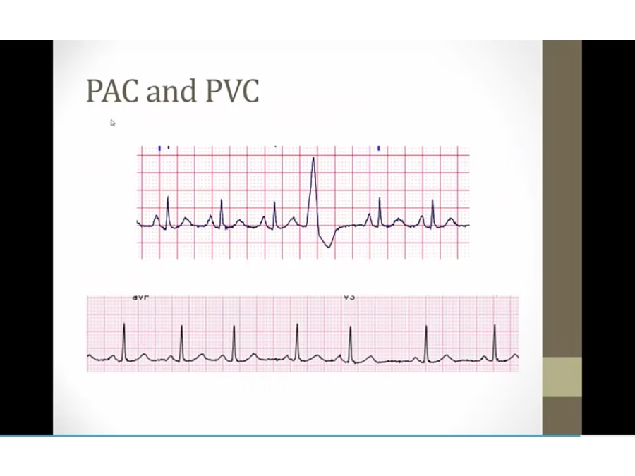
- top: early wide QRS, PVC, something stimulated ventricles to contract on its own (high catecholamines, infection, surgery)
- bottom: early narrow QRS with compensatory pause after
Links to this note