27 Retina
Structures
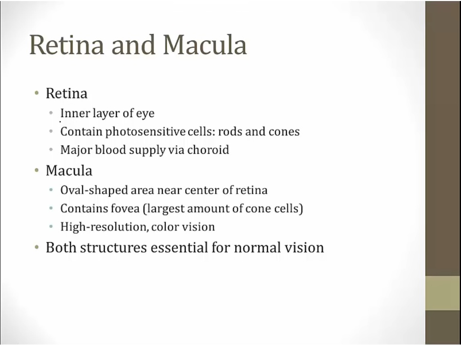
- retina: most important structures. Other structures support its function
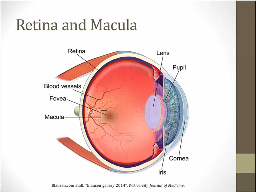
- blue layer: choroid, vasculature
Pathology
Retinitis Pigmentosa
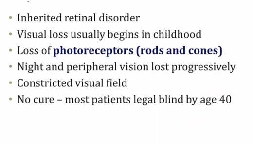
- AD, AR, or X-linked.
- association with abetalipoproteinemia
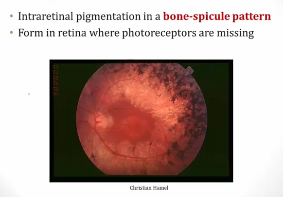
- no real inflammation, just loss of photoreceptors.
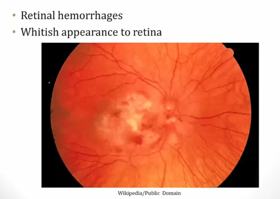
Retinitis
- floaters: dark spots in visual field
Hypertensive retinopathy
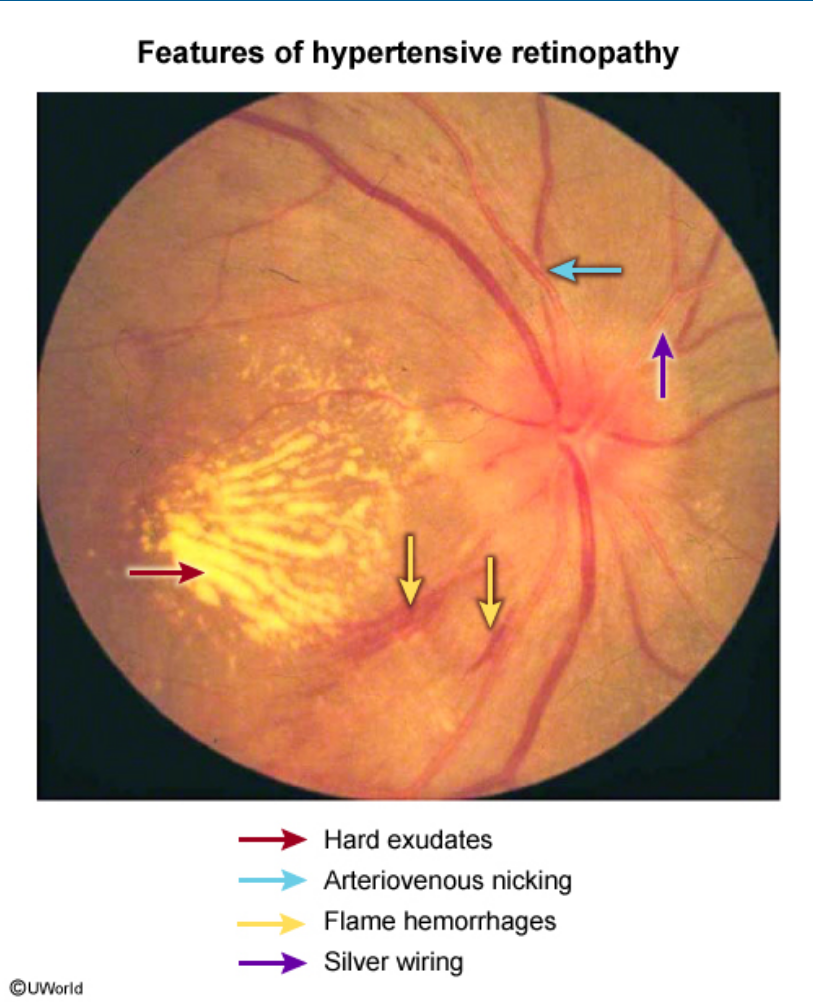
Hypertensive retinopathy can present with gradual, painless, binocular vision loss and headaches, although most patients have no visual symptoms initially. Characteristic features on funduscopic examination include “copper and silver wiring,” hard exudates, flame hemorrhages, arteriovenous nicking, arteriolar narrowing, and ischemic changes (“cotton wool spots”).
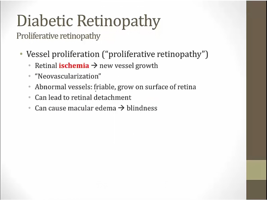
Diabetic Retinopathy
- proliferative vs. nonproliferative
Nonproliferative
- most common type
- microaneurysm: small outpatchings of blood vessels on retina
- cotton wool spot: not unique to diabetes
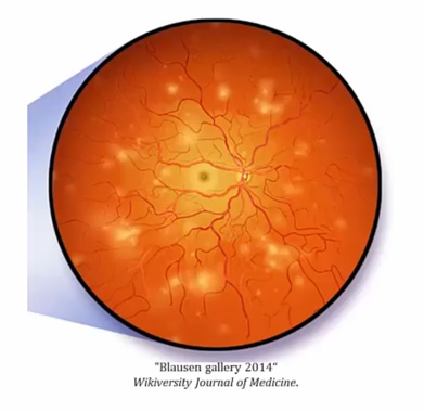
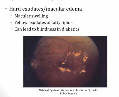
- damage to macula from macular swelling, leads to accumulation of yellow exudates.
- most common form
Proliferative
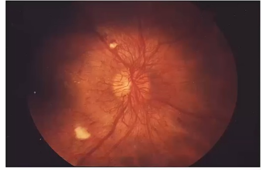
.
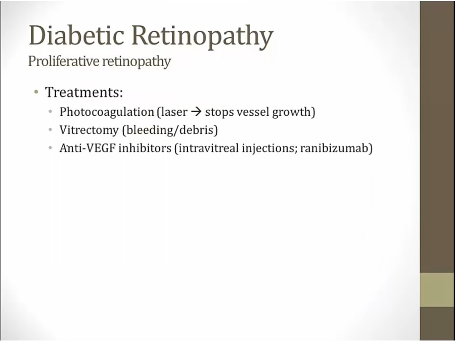
- vitrectomy: removal of part of vitrous humor, done when bleeding from new blood vessels or debris accumulation
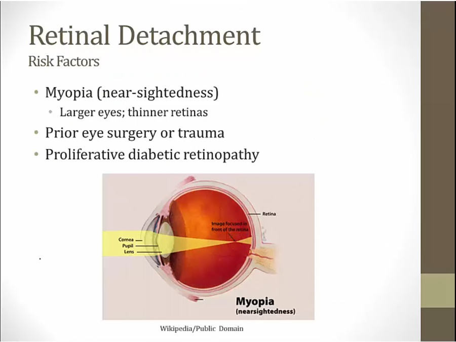
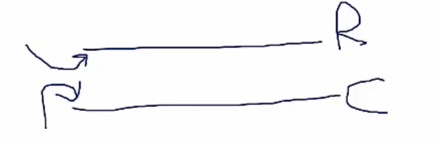
Retinal Detachment
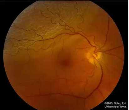
- upper left: retina folded on itself like wrinkled paper.
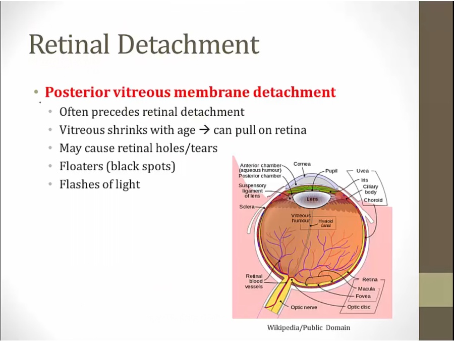
- vitreous humor surrounded by vitreous membrane
- pts complain of floaters, flashes of lights before retina detachment
Amaurosis Fugax
- transient monocular vision loss via emboli that lasts a few seconds.
Retinal Vein Occlusion
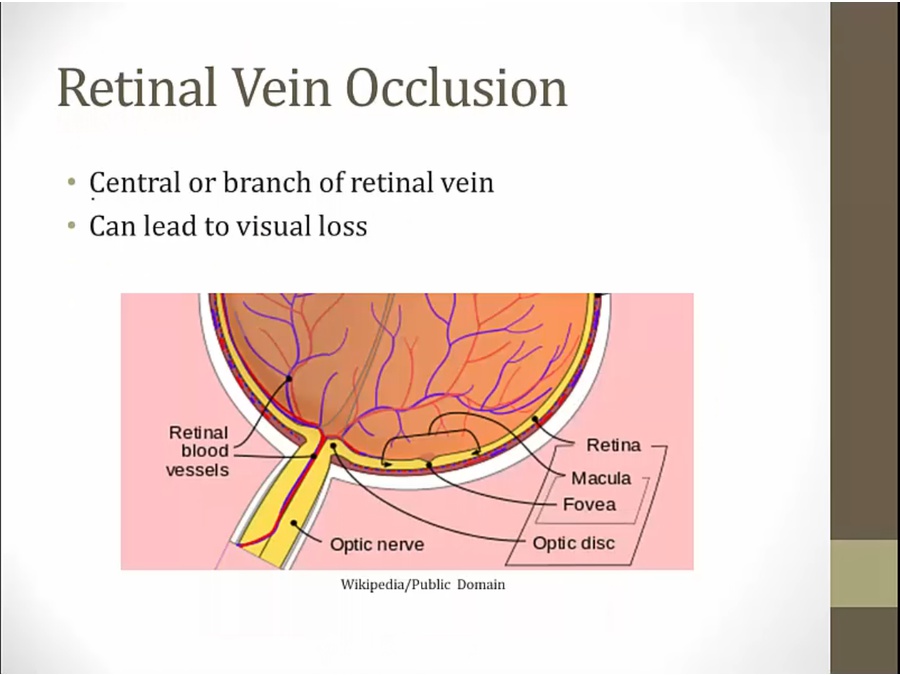
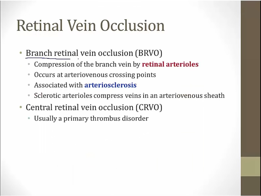
- arteriosclerosis association with hypertension
- arterioles and veins run in AV sheath. Thickened arterioles compress veins
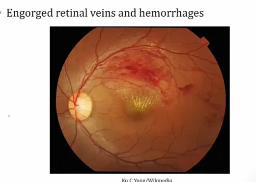
- streaky hemorrhages.
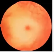
Retinal Artery Occlusion
.
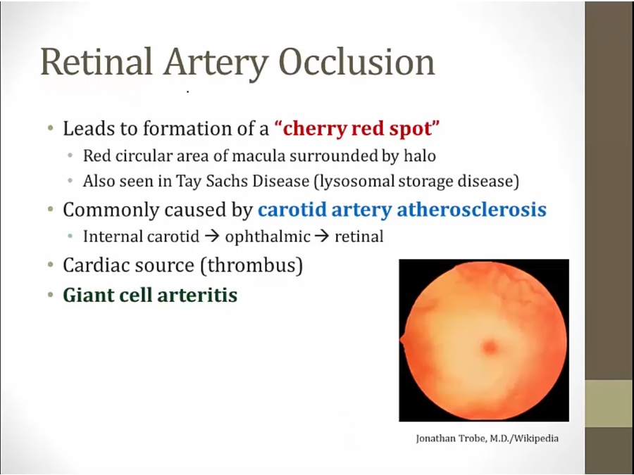
- artery occlusion: ischemia
- Tay Sachs: sphingolipid accumulate around macula
- patient can have painless vision loss
- acute retinal ischemia due to thrombosis or emboli in the central retinal artery (branch of ophthalmic artery). Patients develop sudden-onset, painless loss of vision in one eye, with a cherry red macula and retinal pallor on examination. Additional findings may include a relative afferent pupillary defect (Marcus Gunn pupil).
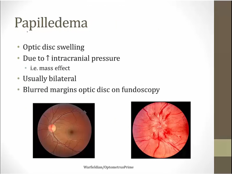
Papilledema
Macula Degeneration
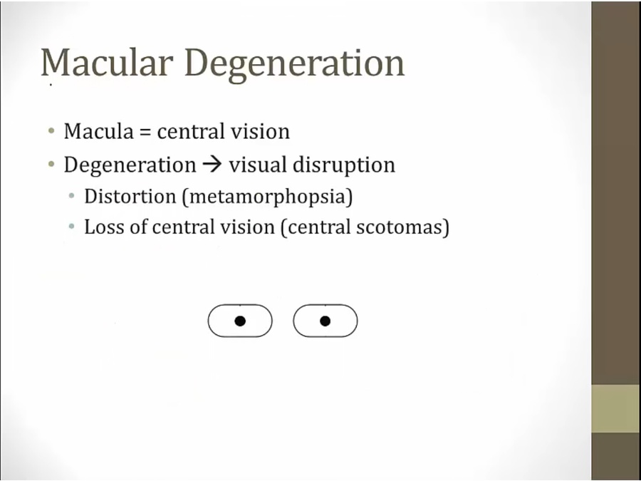
- commonly associated with aging
- macular important for central vision

Dry
- retina, retinal pigment epithelium, bruch’s membrane, choroid:
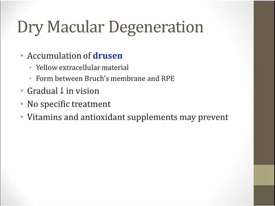
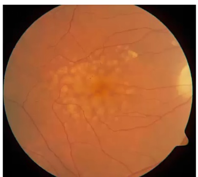
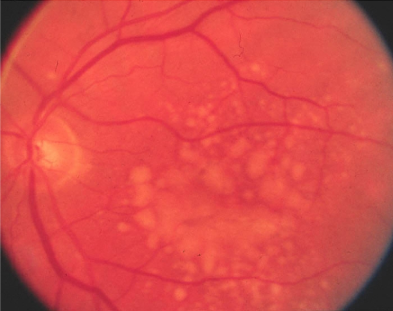
- yellow dots.
Wet
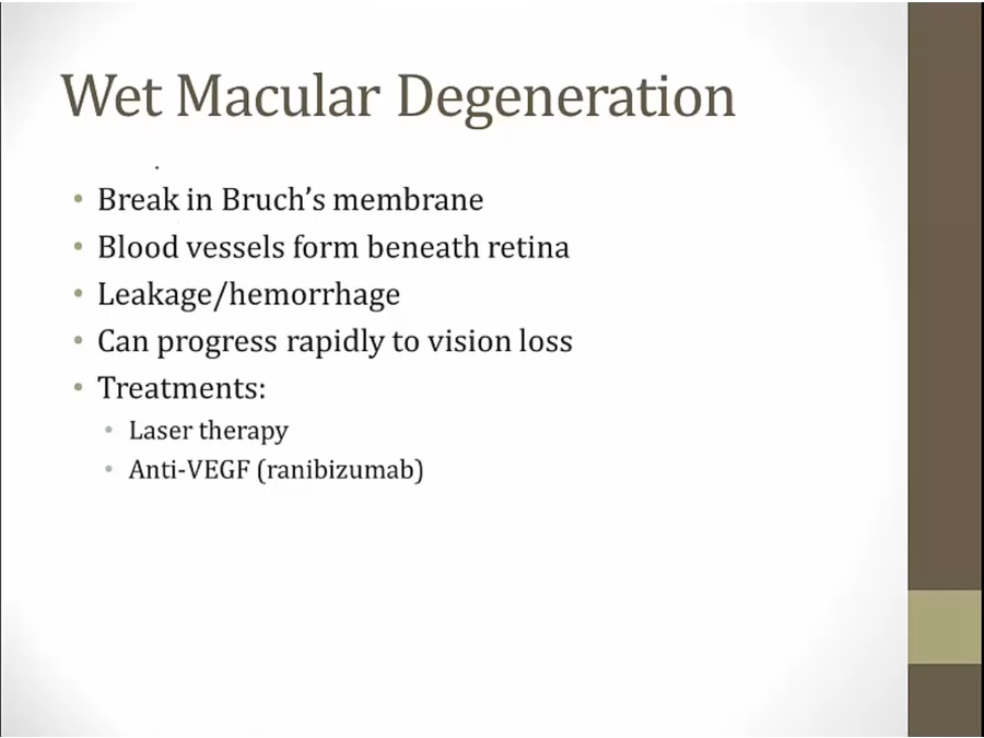
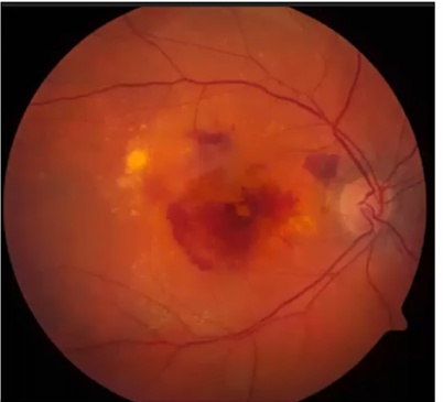
.
- hemorrhage