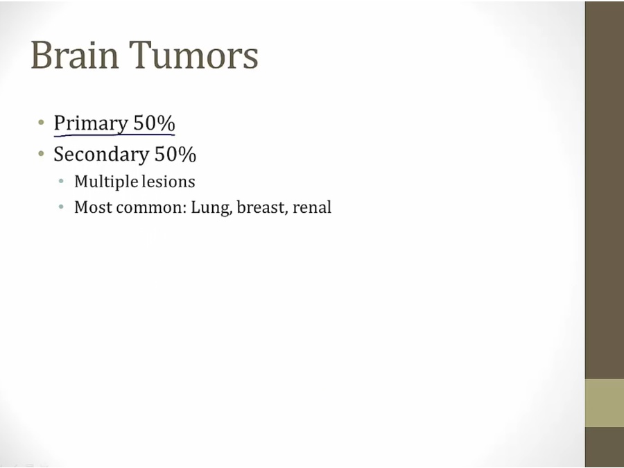
42 Brain Tumors
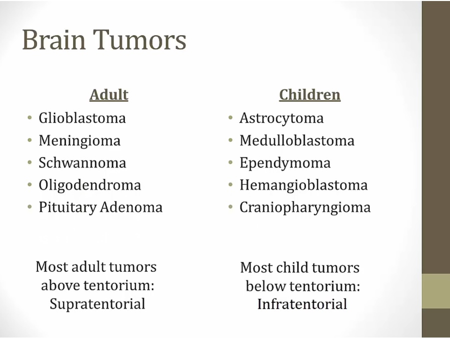
- in order of frequency
- kids: most in posterior fossa near cerebellum
- one of most common symptoms: headache
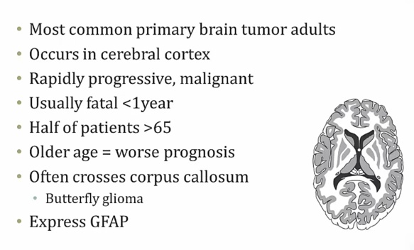
Adult
Glioblastoma
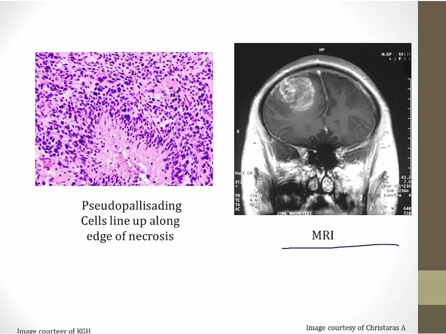
- left: nuclei lining up (schwannoma, nuclei line up on their own)
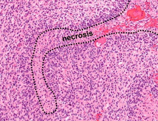
- pseudopallisading: nuclei lining up because being pushed by necrosis from middle
- right: tumor invading cerebral cx.

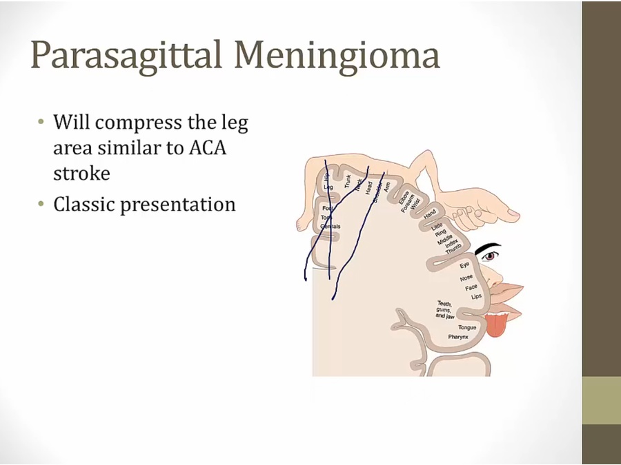
Meningioma
- not in brain tissue itself, can attach to dura with tail
- if large enough: seizures
- tumor expresses estrogen receptors

- Ca deposits


- whorled appearance
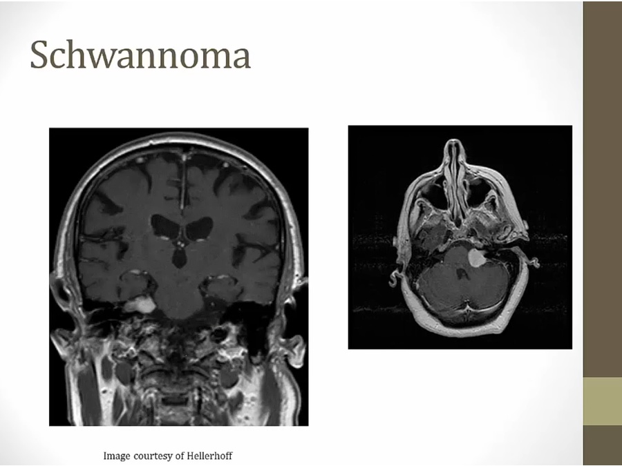
Schwannoma
- locate more peripherally
- S100 marker of schwann cells
Neurofibromatosis
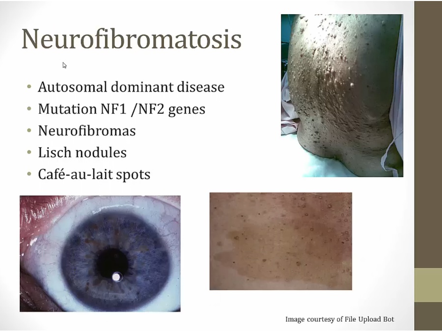
- association with schwannomma
- diffused neurofibromas: upper right, diffused skin nodules all over body
- lower right: cafe au lait, like coffee spilled on skin
- lower left: lisch nodules, brown spots
- neurofibromas: neural crest origin
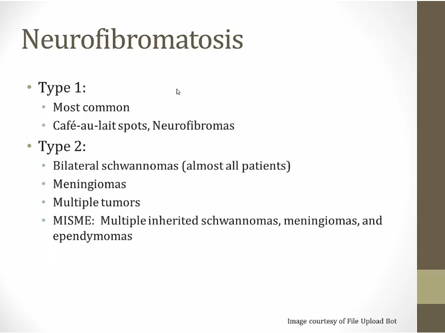
- type 2: almost all get bilateral schwannomas (deaf)
- MISME: multiple tumors
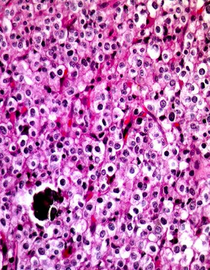
Oligodendroglioma
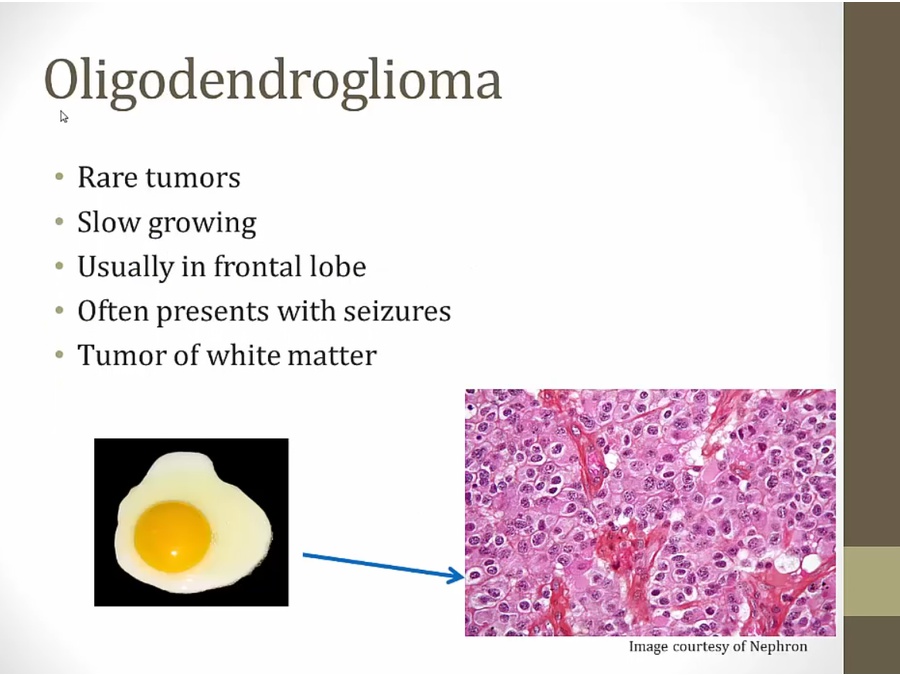
- calcified tumors present in frontal lobe, presenting with seizures
- oligodendrocytes myelinate axons in white matter
- fried egg appearance
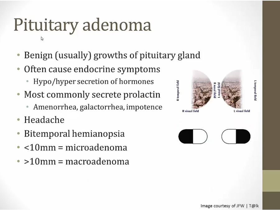
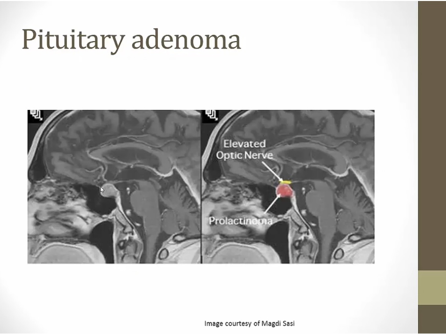
Pituitary Adnoma
Cavernous Hemangioma
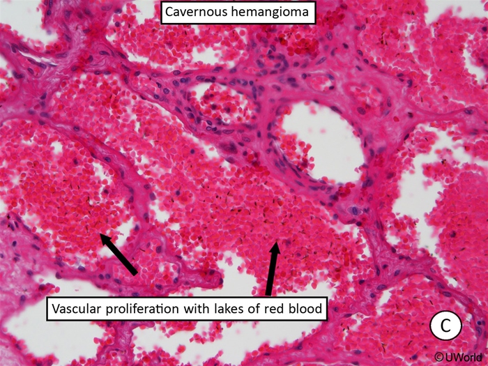
Cavernous hemangiomas are vascular malformations that frequently involve the deeper tissues of the body, such as the liver and brain. Gross examination of hemangiomas reveals a “mulberry-like” appearance due to their purple vascular clusters. Histologically, they are composed of abnormal, dilated blood vessels with a thin adventitia lacking elastic fibers and smooth muscle. The reduced structural support gives them a tendency to leak, causing recurrent hemorrhage.
Most patients with cavernous hemangiomas are asymptomatic, although hemangiomas in the brain may cause neurologic deficits and seizures due to compression of the surrounding tissue and irritation from recurrent bleeding. Surgical resection is indicated for lesions causing intractable epilepsy or progressive neurologic deficits.
Childhood Tumors
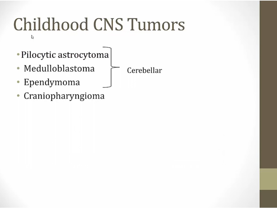
- most common to least common
Pilocytic astrocytoma
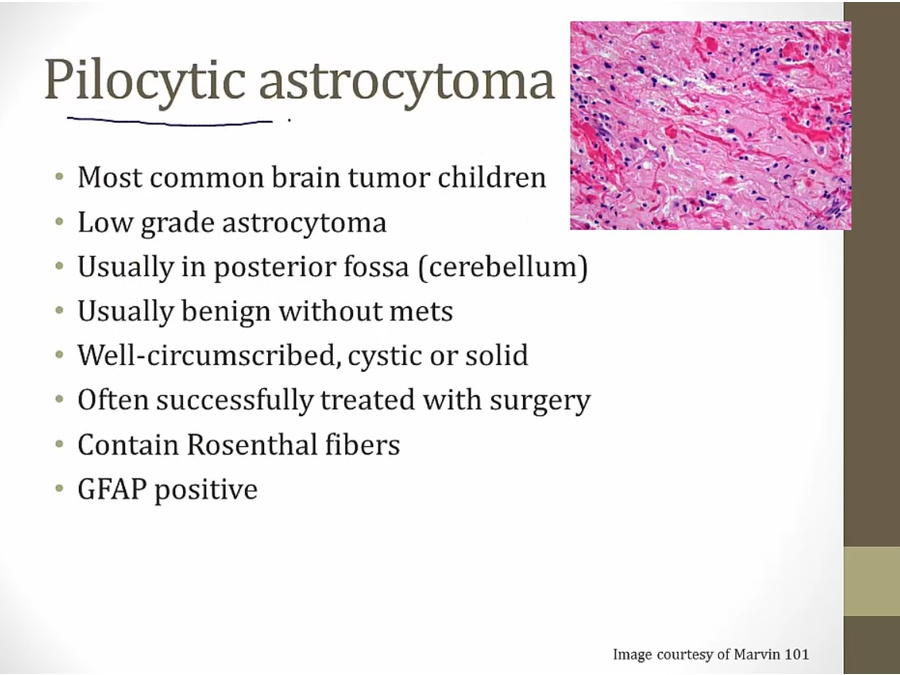
- derived from astrocytes
- cerebellar dysfunction: ataxia, loss of balance
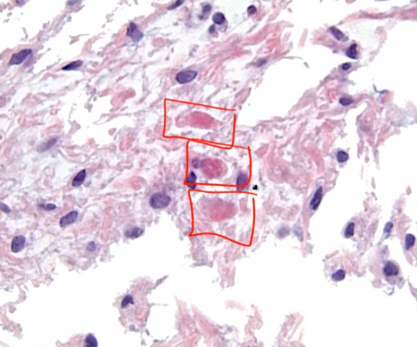
- top right: rosenthal fibers (long red lines), seen anytime gliosis (growth of glial cells)
- GFAP: Glial fibrillary acidic protein, expressed in astrocytes
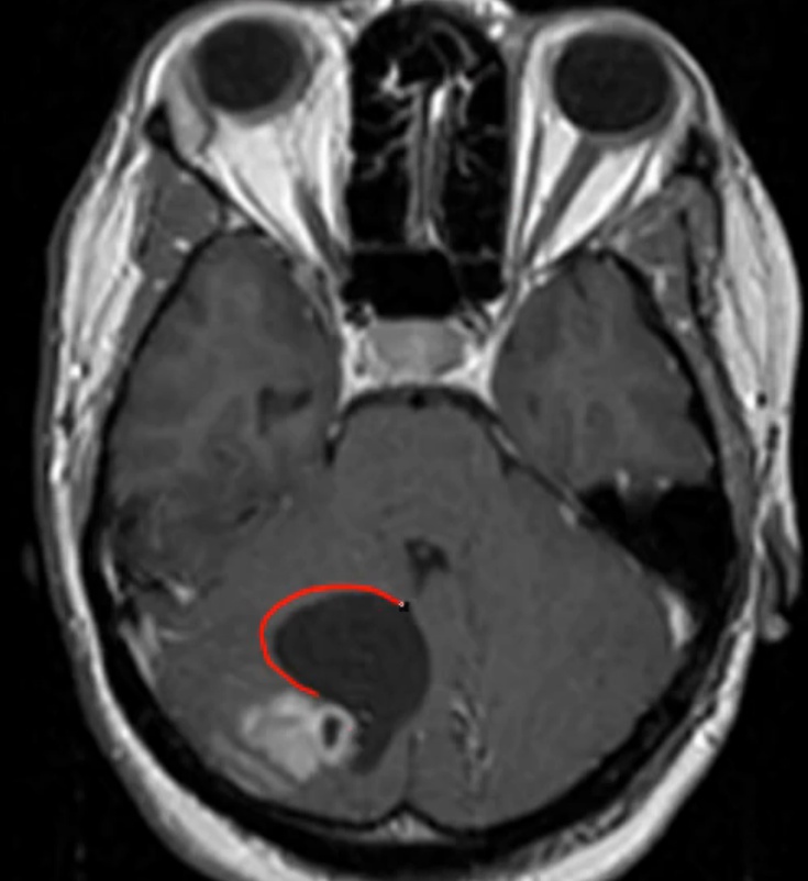
- cystic lesion with nodule growing on its wall
Medulloblastoma
- second most common
- can’t stand/sit up without falling
- neuroectoderm embryonic structures, thus more often in children
- association with Turcot Syndrome
- PNET: tumor suppressor gene
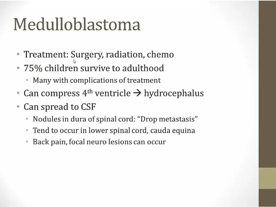
- hearing loss, neurocognitive impairment
- noncommunicating hydrocephalus
- can cause cauda equina syndrome
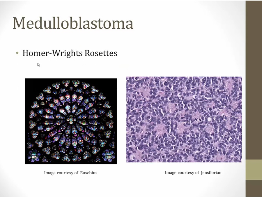
- left: rosettes, circulation orientation of flowers
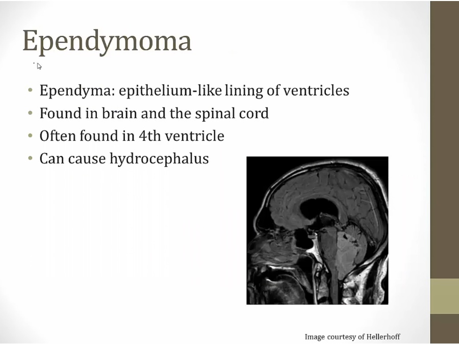
Ependymoma
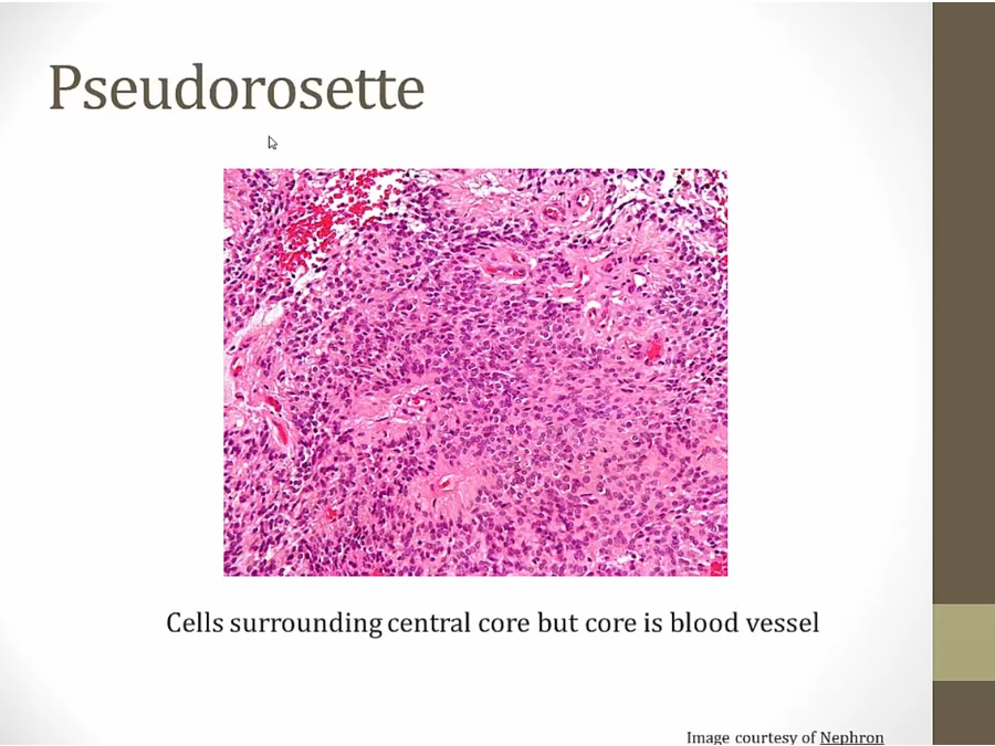
- curcles of cell surrounding blood vessel as central core

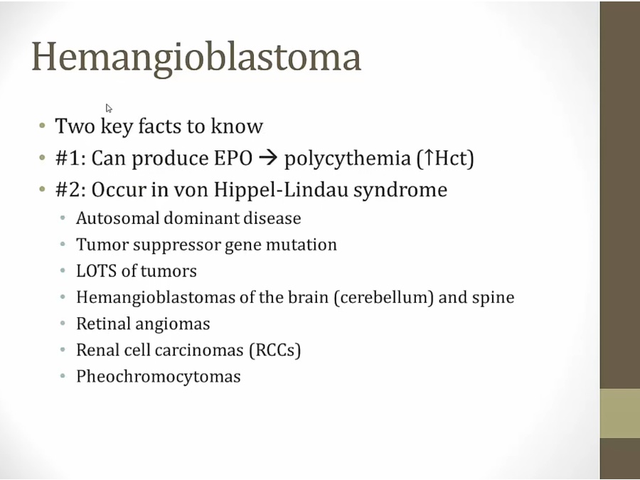
Hemangioblastoma
- EPO
- VHL.
Craniopharyngioma
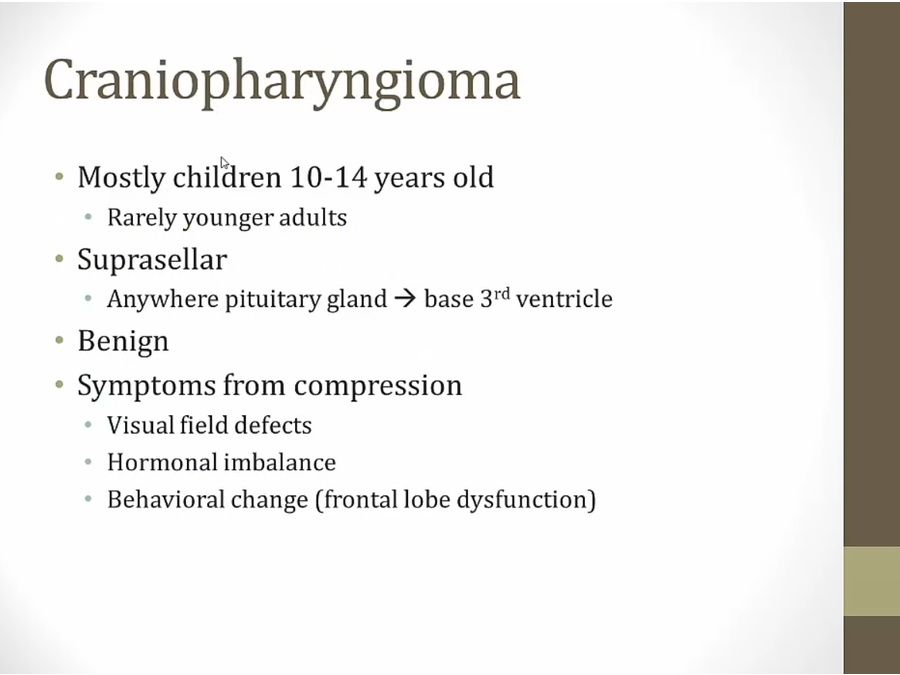
- occur above sella tursica
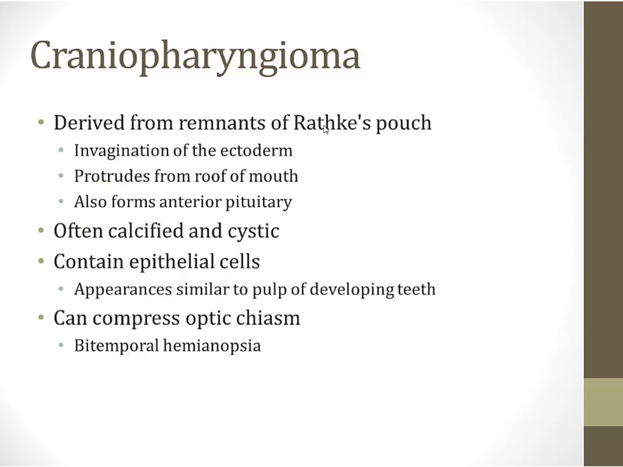
- calcified and look like teeth because derived from mouth tissues
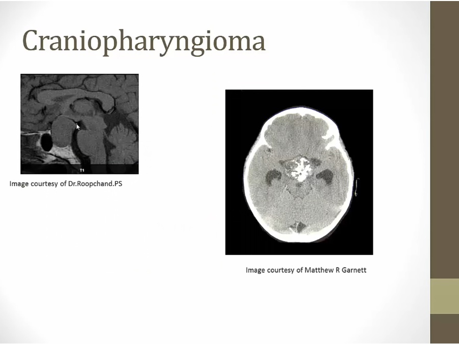
- right: Ca inside
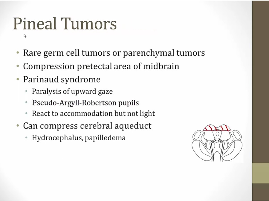
Pineal tumor
Histology and markers
- craniopharyngioma
- meningioma
- glioblastoma, pseudopallisading
- oligodengroglioma, fried egg
- rosenthal fibers, pilocytic astrocytoma
- rosettes, medulloblastoma
- pseudorossettes, ependymoma
- Schwannoma
- craniopharyngioma
- meningioma