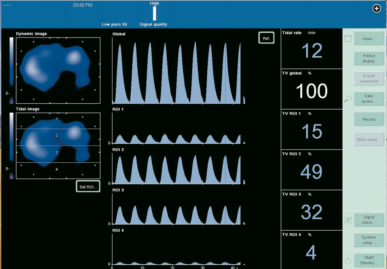
electric impedance tomography can help with avoiding VILI
- related: ICU intensive care unit
- tags: #permanent
- electric impedance tomography (EIT) works by placing sensors around mid chest
- small electrical signals are applied and then received
- air and tissue in the lung returns different impedance
- signals returned then produce cross sectional image of gas volume in the lung
- this can be helpful in preventing VILI: ventilator induced lung injury VILI happens from over distention of normal lung?

- the four quadrants of the lungs are plotted in the picture with tidal volume for each
- regions of interest (ROI) arranged ventral to dorsal. 12
Links to this note
Footnotes
-
Bachmann MC, Morais C, Bugedo G, et al. Electrical impedance tomography in acute respiratory distress syndrome. Crit Care. 2018;22(1):263. PubMed ↩
-
van der Zee P, Somhorst P, Endeman H, et al. Electrical impedance tomography for positive end-expiratory pressure titration in COVID-19-related acute respiratory distress syndrome. Am J Respir Crit Care Med. 2020;202(2):280-284. PubMed ↩