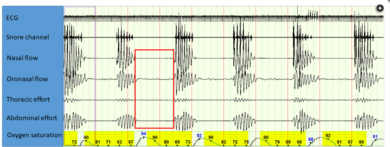
example PSG tracings

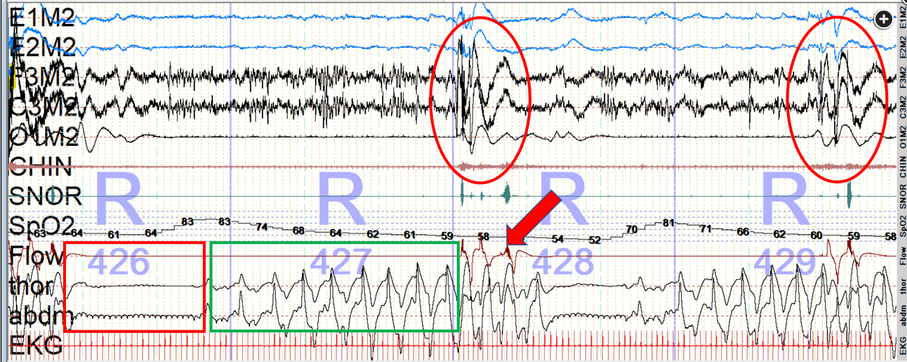
- mixed apneas
- REM sleep 2-min recording. Red box shows central apnea component, green box shows obstructive apnea component, red ovals show cortical arousals, and red arrow shows increase in upper airway dilator muscle activity and patency of the upper airway with resumption of effective flow during cortical arousal. EEG, electro-oculography, and electromyography are necessary in polysomnography to stage sleep into REM and non-REM sleep. Abbreviations: E1M2 and E2M2, referential electro-oculogram recordings from the left eye referred to the right mastoid lead (E1M2) and the right eye referred to the right mastoid lead (E2M2); F3M2, C3M2, and O1M2 represent three referential EEG recordings, from the left frontal, left central, and left occipital areas, respectively; CHIN, electromyographic recording from submentalis region; SNOR, snoring channel; thor, thoracic effort belt; abdm, abdominal effort belt; EKG, ECG.


Links to this note


