histopathology of various lung cancers
- related: lung mass and cancer
- tags: #literature #pulmonology
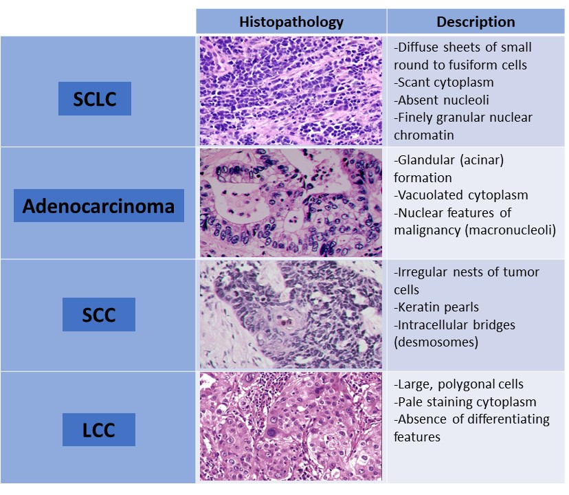
 Histopathologic features of SCLC, adenocarcinoma, SCC, and LCC. Abbreviations: SCLC, small cell lung cancer; SCC,
squamous cell carcinoma; LCC, large cell carcinoma.

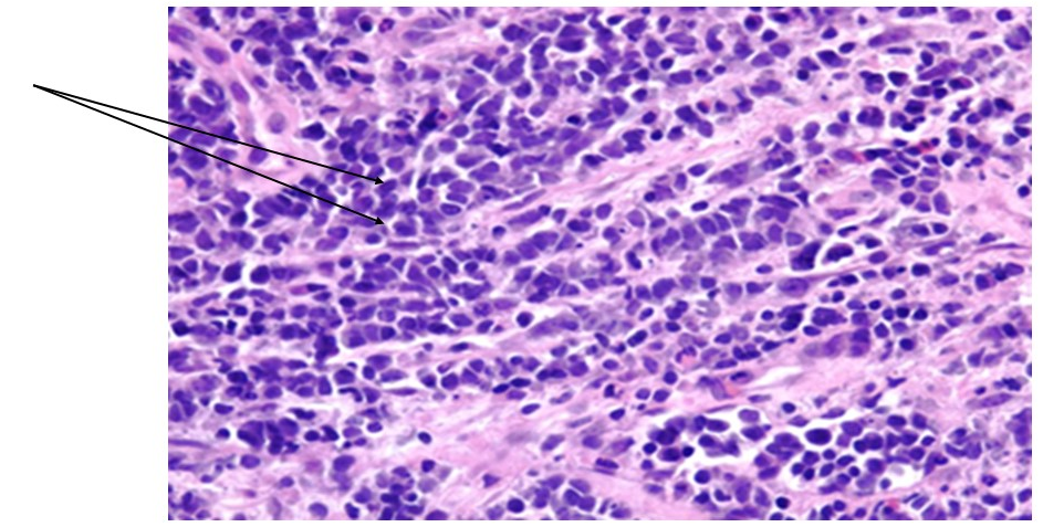
- Histopathologic image of small cell lung cancer showing diffuse sheets of small round to fusiform cells with scant cytoplasm and inconspicuous or absent nucleoli (arrows). Hematoxylin and eosin stain (×40 magnification).
A percutaneous lung biopsy was pursued in this case because the patient had clear evidence of distant metastatic disease, thereby precluding the need for bronchoscopy with endobronchial ultrasonography for staging purposes. Histopathologically, SCLC is defined by the presence of diffuse sheets of small round to fusiform cells with scant cytoplasm and inconspicuous or absent nucleoli with finely granular nuclear chromatin (Figures 3 and 4). Additional nuclear features include molding, smudging, and a high mitotic rate. Immunohistochemical (IHC) markers such as synaptophysin, chromogranin, and CD56 can also be helpful with confirmation of the diagnosis (Figure 5).
Adenocarcinoma is the most common type of lung cancer and accounts for approximately one-half of lung cancer cases. Histologic diagnosis of adenocarcinoma requires evidence of neoplastic gland formation, IHC pneumocyte marker expression, or intracytoplasmic mucin (choice A is incorrect) (Figures 4 and 5). There is significant variation in the extent and architecture of neoplastic gland formation. The major nonmucinous subtypes are acinar, papillary, micropapillary, lepidic, and solid. The World Health Organization classification emphasizes that tissue specimens should be managed not only for pathologic diagnosis but also to preserve tissue for molecular studies, which have important treatment implications, such as use of targeted therapies for certain subsets of patients.
A histological diagnosis of squamous cell carcinoma (SCC) is predicated on the presence of keratinization and/or intercellular bridges (desmosomes) or by IHC findings consistent with SCC (choice B is incorrect) (Figures 4 and 5).
Large cell carcinoma (LCC) constitutes a minority of lung cancer cases. These tumors are devoid of lineage-specific differentiation and lack the morphologic and IHC evidence of adenocarcinoma, SCC, or neuroendocrine carcinoma (such as SCLC) (choice C is incorrect). Therefore, LCC can be a diagnosis of exclusion. The tumor cells are large and polygonal, with prominent pleomorphic nucleoli and abundant pale staining cytoplasm without differentiating features (Figure 4).