PSG tracings for sleep disordered breathing
- related: Sleep and Sleep Disordered Breathing
- tags: #permanent
- Polysomnogram (PSG): includes EEG, ECG, EMG, electooculogram (EOG)
- can diagnose variety of disorders including central sleep apnea
- split night: 1/2 diagnose, 1/2 treatment for OSA
- First 2 hours AHI must be > 15 to continue
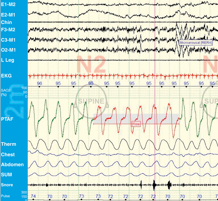
- M1 and M2: EEG signals
- PTAF (pressure transducer): nasal pressure flow signal. Measures pressure difference between inhale and exhale to determine airflow. Better for hypopneas
- Therm or Tflow2: thermositor. Gives similar information as PTAF but uses temperature difference. Better for apnea.1
- Chest/Abdomen: movement of muscle
- snore: snoring2
- This PSG shows example of RERA with airflow obstruction but no desat followed by microarousal
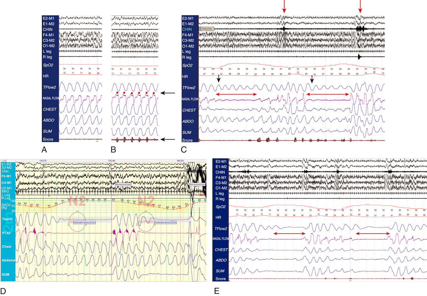
- A: normal breathing
- B: simple snoring
- C: hypopnea: no nasal flow but has thermistor signal
- D: obstructive apnea: both Tflow and nasal flow signal absent. There is muscle movement so obstructive
- E: central apnea: lack of muscle movement shows central apnea
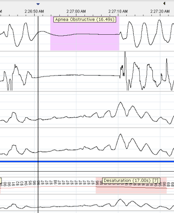
Example of apnea:
Links to this note
-
sleep disordered breathing is characterized by apnea, hyponea, hypoventilation, RERA
- obstructive apnea is when there is respiratory effort in event. PSG tracings for sleep disordered breathing