air bronchogram on ultrasound differentiates between atelectasis vs pneumonia
- related: chest imaging
- tags: #literature #pulmonology
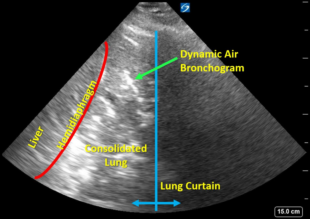
The history in this stem is that of a man who has risk factors for each of the four diagnoses offered: lung cancer, pneumonia, empyema, and pneumothorax. The learner is offered an ultrasonographic video of the right base from which to narrow down the differential diagnosis. A frame shot of the video is shown in Figure 1 and is labeled for the learner’s convenience. Please refer to the video, as the key findings require motion. A lung curtain from the still aerated right upper lobe can be seen moving back and forth from the right side of the video. There is “sonographic hepatization” of the right lower lobe, a term used to describe dense consolidation where the airless lung takes on the ultrasonographic appearance of liver. The short, segmented, echogenic structures within the area of consolidated lung are bronchi that contain minute amounts of air, rendering them hyperechoic. In the video, one or two of these bronchi contain small bubbles of air that are moving back and forth with tidal respiration. These are termed “dynamic air bronchograms.” Lichtenstein and colleagues studied 52 patients with proven bacterial pneumonia and 16 patients with resorptive atelectasis typical of patients with airway occlusion from a tumor. The presence of dynamic air bronchograms had a specificity of 94% and a positive predictive value of 97% for the diagnosis of pneumonia. In other words, the presence of dynamic air bronchograms in this patient is most likely to represent pneumonia rather than atelectasis from central airway obstruction. The sensitivity and negative predictive value of dynamic air bronchograms were lower (61% and 43%, respectively). Thus, the absence of this finding does not exclude a diagnosis of pneumonia.
Empyema in this location might look very complex and echogenic, but it would not demonstrate the characteristic short hyperechoic static and dynamic air bronchograms. If pneumothorax had been present, the interface between the chest wall and the pneumothorax would generate bold A-line artifacts resembling normal lung minus lung sliding.1234
Links to this note
Footnotes
-
Gillman LM, Panebianco N, Alkadi A, et al. The dynamic sonographic air bronchogram: a simple and immediate bedside diagnosis of alveolar consolidation in severe respiratory failure. J Trauma. 2011;70(3):760. PubMed ↩
-
Lichtenstein D, Mezière G, Seitz J. The dynamic air bronchogram. A lung ultrasound sign of alveolar consolidation ruling out atelectasis. Chest. 2009;135(6):1421-1425. PubMed ↩
-
Shah A, Oliva C, Stem C, et al. Application of dynamic air bronchograms on lung ultrasound to diagnose pneumonia in undifferentiated respiratory distress. Respir Med Case Rep. 2022;39:101706. PubMed ↩