azygos lobe is normal anatomic variant
- related: chest imaging
- tags: #literature #pulmonology
- azygos vein is displaced laterally, creating pleural fissure in the apical segment of right upper lobe
- not true lung lobe because it doesn’t have its own bronchus
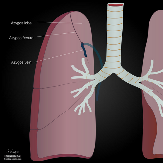
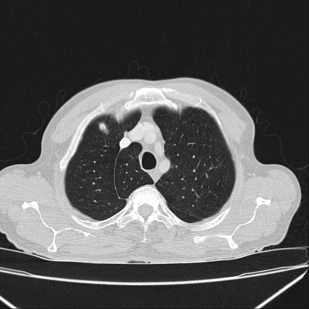
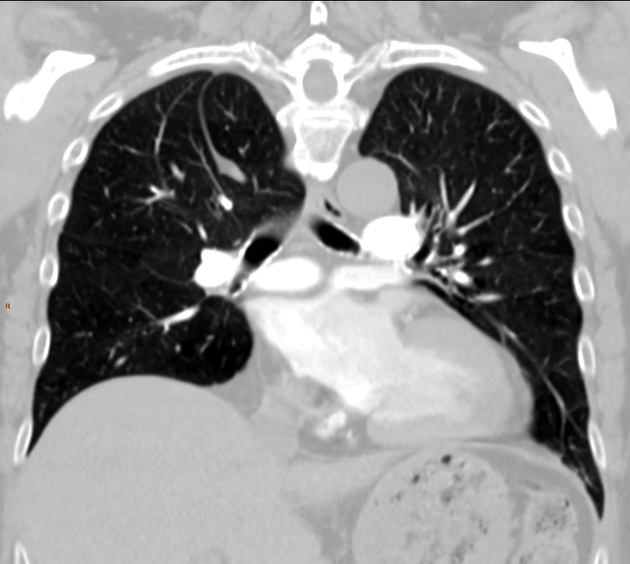
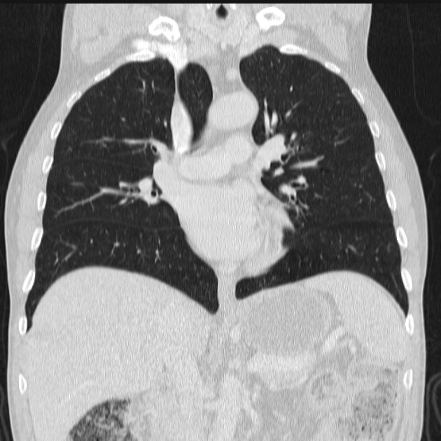
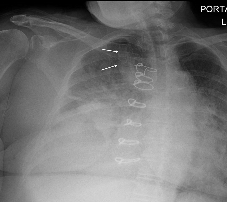
Normal anatomic variation is important for the intensivist to recognize so that findings are not confused for pathology. This chest radiograph shows an azygos fissure—the most common accessory fissure, present in roughly 0.5% of patients. The azygos fissure and lobe form during embryology, when the posterior cardinal vein (precursor of a segment of the azygos vein) aberrantly migrates through the upper lobe. As a result, two layers of pleura are carried through the lobe to produce the azygos fissure. This can be confused with abscess, bulla, or pneumothorax. The structure is even more readily evident on CT scanning (Figure 2). An azygos lobe is not a true lobe because it lacks a bronchus. There is no medical significance to an azygos fissure, but it does require awareness by an operating surgeon.
Retained guidewires are common but are more radiopaque than the finding seen here. Pneumothorax will generally not leave evident lung distal to the pleural line, as is seen here. Moreover, the location on this chest radiograph is typical of an azygos lobe. Colonic and gastric interposition used in treatment of esophageal cancer can produce confusing shadows on a chest radiograph, but the appearance here is not typical.234
Links to this note
Footnotes
-
Al-Mnayyis A, Al-Alami Z, Altamimi N, Alawneh KZ, Aleshawi A. Azygos lobe: prevalence of an anatomical variant and its recognition among postgraduate physicians. Diagnostics (Basel). 2020;10(7):470. PubMed ↩
-
Tran CT, Miao KH, Lui F. Anatomy, thorax, lung azygos lobe. In: StatPearls. Treasure Island (FL): StatPearls Publishing; July 24, 2023. PubMed ↩