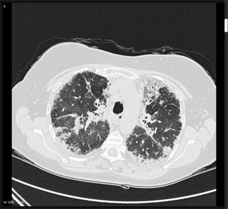
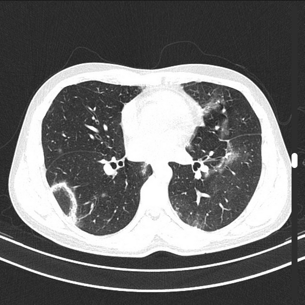
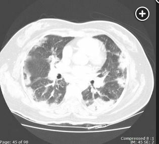
chronic eosinophilic pneumonia has consolidation with peripheral predominance
- related: acute and chronic eosinophilic pneumonia AEP and CEP
- tags: #permanent
- predominance for upper and middle lobes 1


- Bilateral in 50% of case on normal CT and up to 97% on HRCT
- upper lobe predominant, lower lung involvement is infrequent
- pleural effusions are infrequent unlike AEP [^2]
- Chest imaging findings are that of bilateral, usually peripheral or pleural-based, opacities and are often described as the “photographic negative” of pulmonary edema (Figure 1), and, although characteristic, this finding is usually seen in only about 25% of patients.