An Algorithmic Approach to Lung Disease
- related: Pulmonary Diseases, Mariencheck radiology conference composite
- tags: #literature
- source: algorithmic approach
Steps for initial review
- initial review of CXR, including scout film
- review of data acquisition and reconstruction
- initial scrolling of CT to establish quality and overview of disease
- determine category of disease
- reticular
- nodular
- diffuse alteration in density
2. Data Acquisition and Reconstruction
- standard technique includes 1-2.5 mm edge enhanced, sharp reconstruction
- expiratory imaging evaluates air trapping
- prone image differentiates dependent atelectasis vs early interstitial disease
- MIP (maximum intensity projection) image for detecting small nodules
3. Scrolling to Establish Overview
- assess overall lung volumes, motion artifact, inspiratory efforts
- figure out major abnormal pattern
5. Reticulation
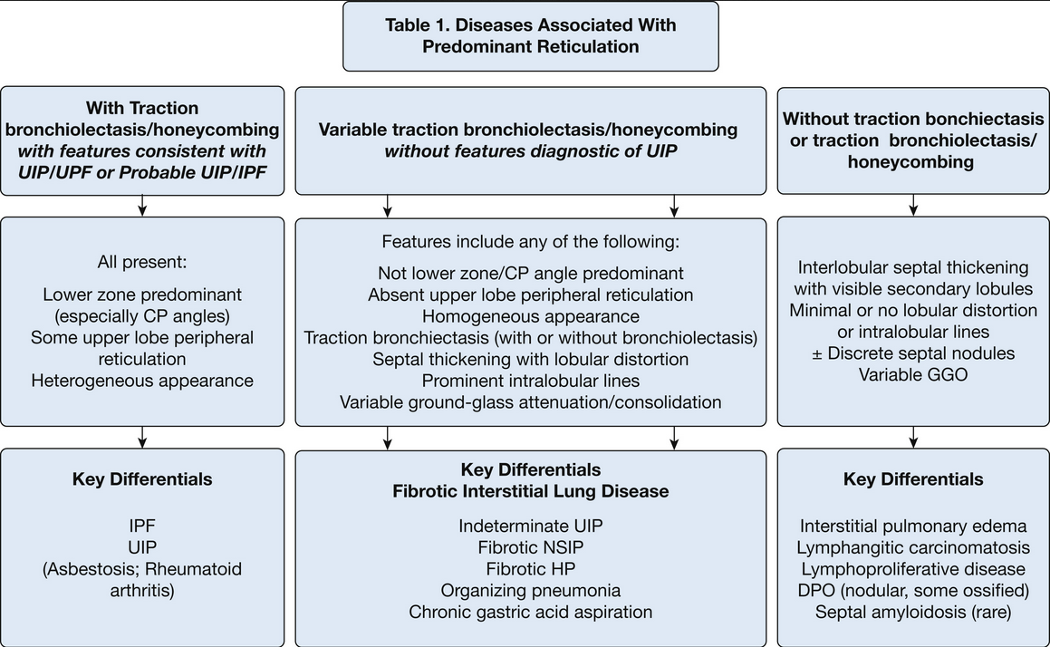
- criteria for radiographic diagnosis of UIP includes lower lobe honeycombing and bronchiectasis
- peripheral reticulation with distortion and angulation of secondary lobule
- subpleural and costophrenic angle predominance with component of upper lobe
- nonsegmental distribution (crosses fissures)
- traction bronchiectasis or honeycombing
- heterogenous appearance
- Probable UIP
- has characteristics of underlying fibrosis
- NSIP associated with systemic sclerosis. NSIP has shared imaging features with UIP
- chronic fibrotic HP
- Interlobular septal thickening
- dendriform pulmonary ossification can occur more in the posterior and lateral basilar segments of lower lobe, associated with recurrent gastric aspiration in elderly men
6. Nodular Diseases
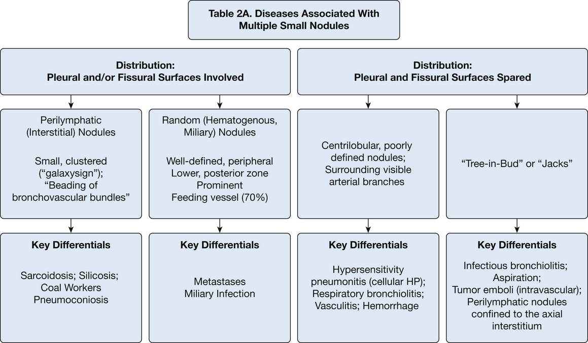
- perilymphatic distribution is characterized by small clusters of nodules along more proximal distribution along fissures or pleura.
- pulmonary sarcoidosis findings include mediastinal lymphadenopathy and perilymphatic nodules
- miliary TB finding includes diffuse nodules in random distribution
- hypersensitivity pneumonitis can have centrilobular nodules with air bronchogram formation
- smoking related respiratory bronchiolitis can have centrilobular nodules with more upper lobe distribution
- tree in bud opacities are associated with nonspecific infections
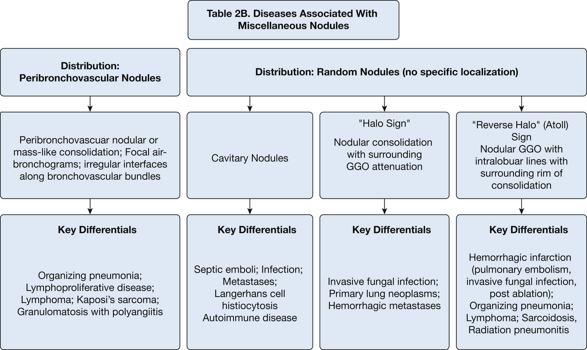
- septic lung emboli are cavitary nodules with peripheral, perivascular, and lower lobe predominance
- halo sign is nodular consolidation with surrounding ground glass
- reverse halo sign has core ground glass and surrounding consolidation
7. Diffuse Alteration in Density
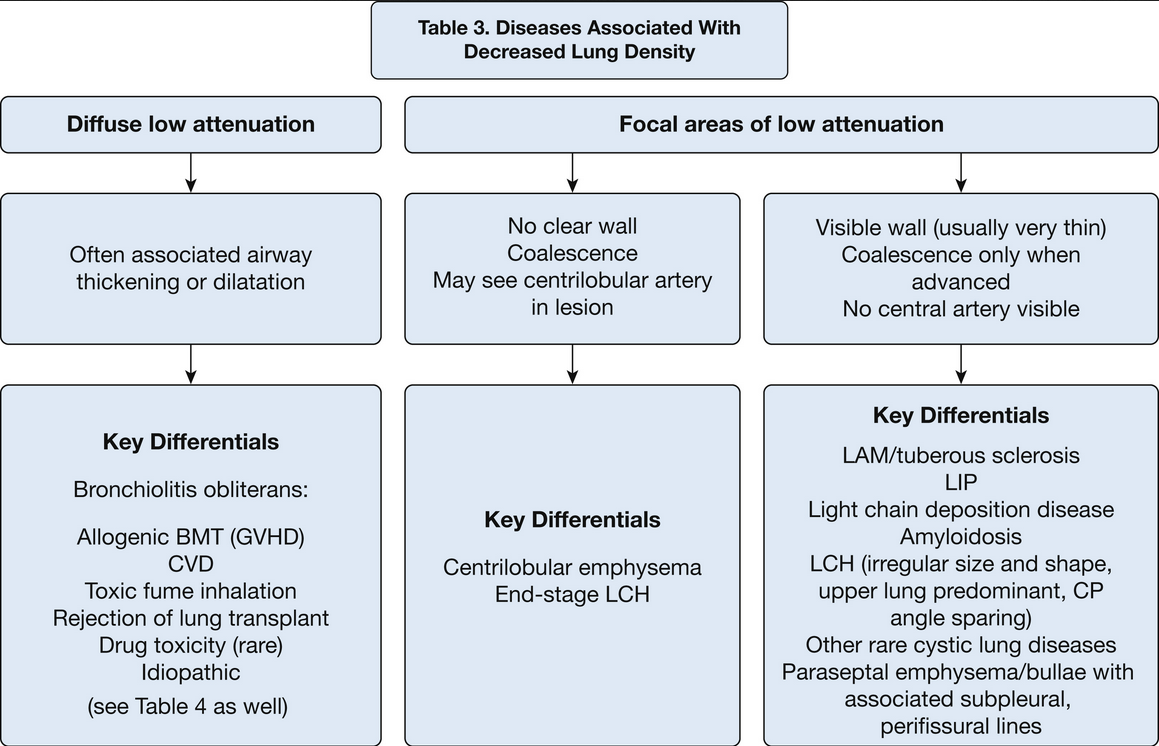
- small airway disease with air trapping
- focal disease without air trapping
- diffuse cystic disease
- lymphangioleiomyomatosis has cystic lesions with chylous effusions on chest imaging
- langerhans histiocytosis has cysts in bizarre groups and upper zone predominant
- amyloidosis have cystic lung lesions in subpleural and perifissure regions
- lymphoid interstitial pneumonia LIP can have ground glass opacities with peribronchovascular cystic lesions, differentiate it from NSIP
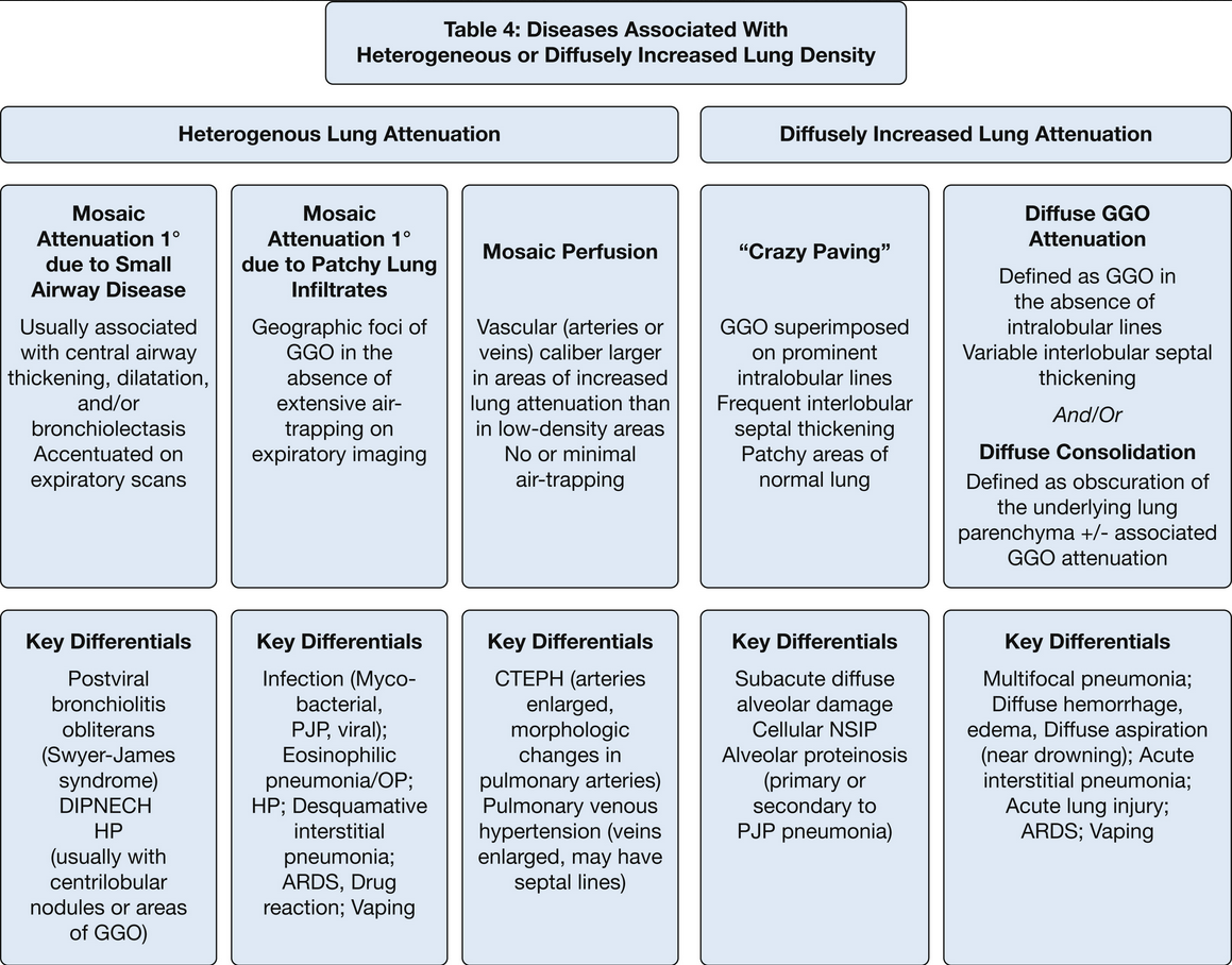
- image findings in hypersensitivity pneumonitis include GGO, centrilobular micronodules, and mosaicism
- CTEPH has mosaic findings with large vessels in denser lung areas
- pulmonary hemorrhage has ground glass opacity on imaging
- alveolar proteinosis have crazy paving patterns
- acute eosinophilic pneumonia has ground glass opacitiees with consolidation on CT imaging
- chronic eosinophilic pneumonia has consolidation with peripheral predominance
Links to this note
-
- [^1]: SEEK Questionnaires
-
lymphangioleiomyomatosis has cystic lesions with chylous effusions on chest imaging
-
bronchiolitis with air trapping can have appearance of decreased lung density on expiration
-
amyloidosis have cystic lung lesions in subpleural and perifissure regions
-
septic lung emboli are cavitary nodules with peripheral, perivascular, and lower lobe predominance
-
CTEPH has mosaic findings with large vessels in denser lung areas
-
halo sign is nodular consolidation with surrounding ground glass
-
langerhans histiocytosis has cysts in bizarre groups and upper zone predominant
-
reverse halo sign has core ground glass and surrounding consolidation
-
tree in bud opacities are associated with nonspecific infections