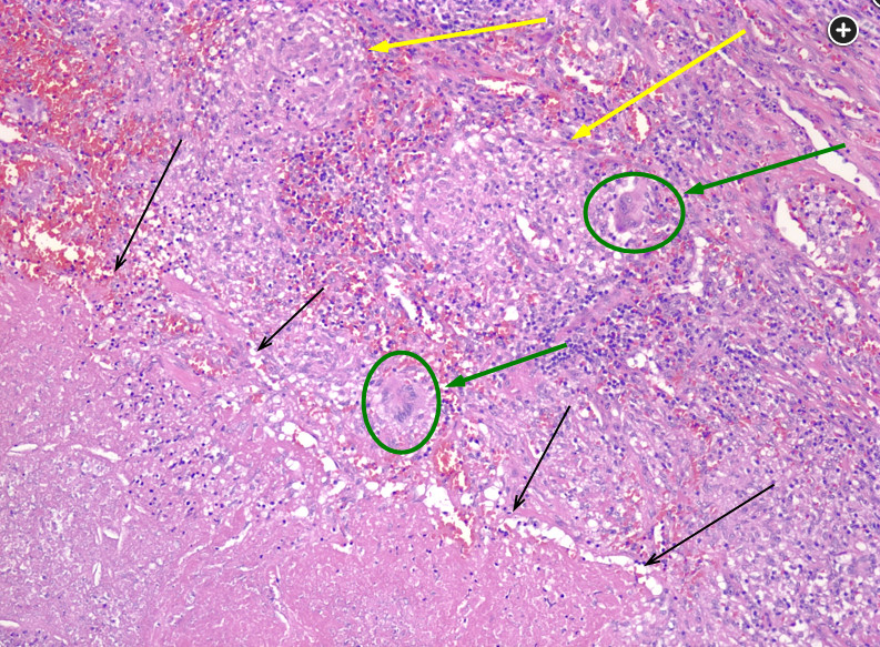
granulomatosis with polyangiitis histology shows necrotizing granulomas
- related: Wegener GPA granulomatosis with polyangitis
- tags: #literature #pulmonology
 The black arrows form a line of necrotizing inflammatory change. Yellow arrows point to nonnecrotizing granulomas. The green arrows point to giant cells.
The third option shows the presence of necrotizing and nonnecrotizing granulomas. Necrotizing granulomatous inflammation is often caused by infections, including mycobacterial infection, and nonnecrotizing granulomas are often associated with certain inflammatory conditions such as sarcoidosis. This photomicrograph (Figure 7) shows areas of nonnecrotizing granulomas (yellow arrows), necrotizing inflammation (in the region indicated by black arrows), as well as giant cells (green arrows) but does not reflect what would be expected for a patient with the radiographic and clinical history provided (eg, the case subject had a negative interferon-γ release assay), which is most congruent with silicosis. It is, however, important to recall that mycobacterial infections are closely associated with underlying silicosis and should be considered clinically as part of the evaluation process.
Granulomatosis with polyangiitis (previously referred to as Wegener granulomatosis) is typically associated with large areas of necrosis with irregular borders (so-called geographic necrosis). These areas of necrosis often take place in an inflammatory background, as shown in Figure 7. However, the history and radiographic features demonstrated in this case are not that of a patient with granulomatosis with polyangiitis.1
Links to this note
-
GPA granulomatosis with polyangitis symptoms include cavitary lung disease and neuropathy
- This patient has a chronic multiorgan system disease characterized by cavitary lung disease (GPA granulomatosis with polyangitis CT chest shows cavitary diseases), peripheral neuropathy, and brain and liver lesions. He has had an extensive workup for infectious and neoplastic diseases, which has been negative. The combination of chronic cavitary lung disease and peripheral neuropathy should raise the suspicion of systemic vasculitis, especially granulomatosis and polyangiitis (GPA). GPA is a necrotizing granulomatous vasculitis (granulomatosis with polyangiitis histology shows necrotizing granulomas) that predominantly affects small vessels of the upper respiratory tract, the lower respiratory tract, and the kidneys, although any organ can be involved.