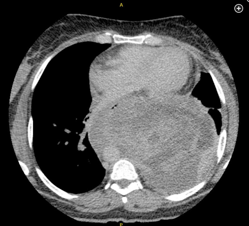
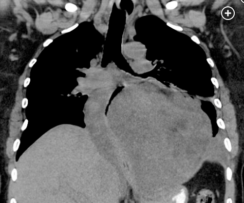
mediastinal liposarcoma is hetergenous mass with adipose tissues
- related: lung mass and cancer
- tags: #literature #pulmonology
This patient has a mediastinal liposarcoma (ML) (choice A is correct). Although liposarcomas are one of the most common soft-tissue malignancies in adults, they are typically found in the retroperitoneum and extremities. Primary ML is considered rare, although it has been observed in most mediastinal compartments, with a variety of histological types. Well-differentiated tumors appear to be the most common and typically grow slowly and along tissue planes, producing symptoms only when they are large enough to compress local structures, as was the case here. Metastatic potential of this histological grade is generally low. CT scans reveal a heterogeneous mass with adipose tissue attenuation (Figures 2 and 3), with a lesser amount of fat seen in the higher grades of ML.


PET scans can be used to distinguish high-grade sarcomas from benign lesions but are considered less useful in lower-grade sarcomas and in general are not recommended as part of the initial workup. Core needle or surgical biopsy is necessary to make the diagnosis. Primary treatment is complete surgical resection. In patients with well-differentiated liposarcomas, anatomical location is critical, with a good prognosis for those whose tumor can be resected with clear margins. Many, however, present with tumor wrapped around vital structures, making it difficult to obtain negative margins, with a high likelihood of subsequent local recurrence. Tumors do not metastasize unless they dedifferentiate, which is associated with a poor prognosis. The efficacy of radiation therapy and adjuvant chemotherapy is unclear.1