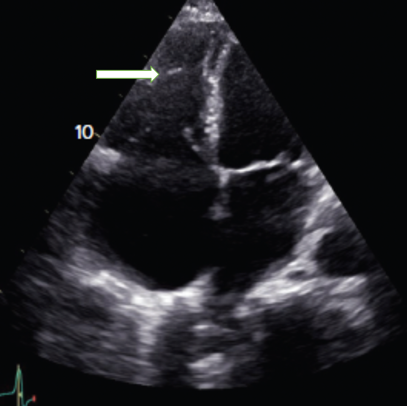
moderator band in RV on ultrasound
- related: cardiac ultrasound essentials
- tags: #literature
 This is an apical four-chamber view of the heart. The arrow is indicating a moderator band (MB) in the right ventricle (RV). An MB is a muscular trabeculation that contains the right bundle branch. It serves as an anchoring structure for the tricuspid papillary muscle (choice A is correct). It extends from the base of the anterior papillary muscle to the ventricular septum. As a prominent trabeculation within the RV, the MB has been implicated in several clinical conditions. It has been suggested that the MB can act as a protective mechanism to resist the overdistension of the RV. In cardiac echocardiography, the MB can be useful as a landmark for the RV, particularly in a congenitally abnormal heart; however, at times, it has also been mistaken for an apical thrombus. It is not usually visible in an adult normal RV given the angles with which the RV is evaluated. It is seen when the RV is dilated and the angle of interrogation changes.
The MB can be seen in any disease state in which the RV is dilated. This includes acute RV infarct, pulmonary embolism, and pulmonary hypertension. It is not 100% specific for a pulmonary embolism (choice B is incorrect). See Video 1 for an example of a dilated RV from pulmonary embolism. The MB is not a thrombus and is a normal structure in the RV. Its presence alone is not an indication for anticoagulation (choice C is incorrect). See Video 2 for an example of a dilated RV with a thrombus. See Video 3 for an example of a clot in transit. If there is a pulmonary embolism found as the reason for RV dilatation, then anticoagulation may be indicated. The MB is an anatomical structure in the RV and not an artifact (choice D is incorrect).1