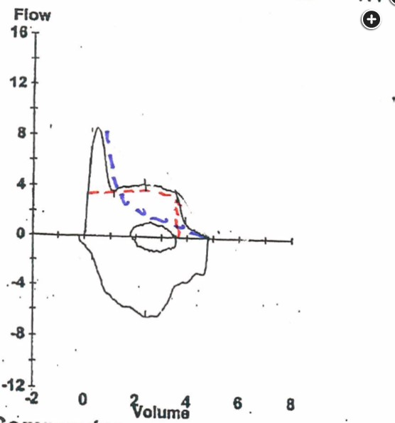
PFT example of lung cancer causing fixed airway obstruction
- related: lung mass and cancer
- tags: #literature #pulmonology

- Inspiratory and expiratory flow volume loop with the conceptual contributions of the two mainstem bronchi. The red tracing indicates a fixed intrathoracic airway obstruction in one bronchus; the blue tracing indicates a typical COPD pattern in the other.

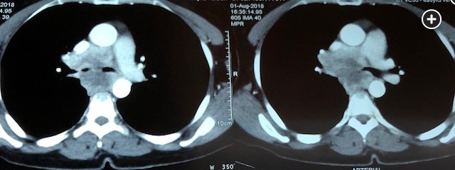
- Chest CT scan illustrates major obstructive lesion in right mainstem bronchus.
The determinants of the forced expiratory flow volume pattern change during the expiratory maneuver. At the beginning, effort is important, and the point of maximal airflow resistance is in the larger airways (4th-8th generation of airways). As exhalation continues, elastic recoil becomes the major source of expiratory pressure (“effort independence”), and the point of maximal airflow resistance moves distally to more collapsible smaller airways. Importantly, a large upper airway obstruction (eg, a large airway mass) produces a fixed resistance that persists throughout exhalation.
The flow-volume pattern in this patient is unusual and, in fact, features the simultaneous effects of moderate COPD in one lung and a large fixed major airway obstruction in the other lung. This can be depicted by overlapping two conceptual flow volume patterns, one from each lung (Figure 2). This was confirmed in this patient with a chest CT scan showing a large endobronchial mass in the right mainstem bronchus (Figure 3) (choice D is correct; choices A, B, and C are incorrect). Interestingly, a similar phenomenon has been noted in unilateral lung transplant patients with an obstructed anastomotic site.1