pulmonary langerhans PLCH has S100 and CD1a positive staining
- related: pulmonary langerhans histiocytosis PLCH
- tags: #literature #pulmonology
On histologic examination, the nodules of PLCH have a stellate appearance and are centered on the small airways. Cysts are formed at the periphery of the nodules by traction on the surrounding alveolar walls or central terminal airway, resulting in variably shaped spaces, typically lacking distinctive lining cells (Figure 5). Langerhans cells have distinctive IHC staining for S-100 protein and are CD1a positive.1
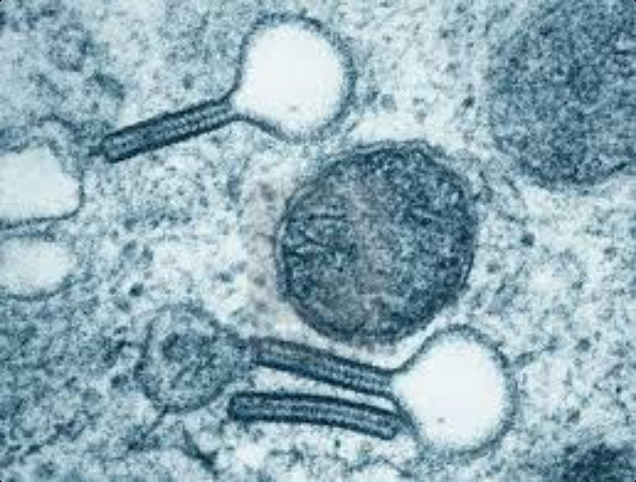
Langerhans cells possess large cytoplasmic “tennis-racket” Birbeck granules.2