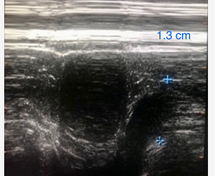
use ultrasound to reveal impaired diaphragmatic excursion
- related: Pulmonology
- tags: #literature #pulmonology
Figure 3 shows a bedside ultrasonographic image revealing impaired diaphragmatic excursion on both right and left sides of 1.5 cm and 1.3 cm, respectively (normal value, 3-8 cm). The diaphragm thickness measured in the zone of apposition at the end of expirations was only 1.2 mm (normal value, 1.8 or greater). The negative inspiratory force was reduced at −30 cm H2O (normal value, −80 to −100 cm H2O).

Bedside ultrasonographic image of the left hemidiaphragm at the zone of apposition during a forced inspiration revealed severely reduced diaphragmatic excursions with a maximum diaphragmatic movement of 1.3 cm.