Warfarin can cause tracheobronchial tree calcification
- related: Pulmonology
- tags: #literature #pulmonology
- 1.chronic warfarin use, ESRD, age can cause 2.tracheobronchial calcification::airway abnormality


source
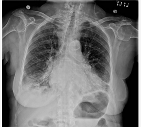
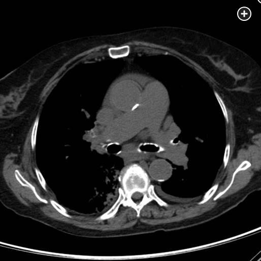
The chest radiograph and chest CT scan reveal prominent calcification of this patient’s tracheobronchial tree. In addition to an association with advanced age and dialysis-dependent chronic kidney disease, this abnormality is associated with chronic anticoagulation with warfarin (choice A is correct). Matrix G1a protein is a vitamin K-dependent inhibitor of medial arterial calcification whose synthesis and activity are blocked by warfarin; this is thought to be the primary cause of this radiologic abnormality. Calcium and phosphorus levels are typically normal in patients exhibiting this radiologic finding. It is seen more commonly in women than men for unclear reasons. Warfarin-associated airway calcification is most commonly seen in older adults, but it has been described in pediatric patients who have required continuous anticoagulation over several years. Vascular calcification can also occur in these patients and prominent aortic and coronary artery calcification are present on this patient’s studies. This prominent radiologic abnormality has no pathophysiologic consequence and does not produce airflow obstruction. It does not require that the warfarin be stopped or that the patient necessarily be switched to an alternative anticoagulant. In this patient, the cause of her progressive dyspnea was ultimately attributed to further decompensation of cardiac function.