bladder uterus FAST exam
- related: ICU intensive care unit
- tags: #literature #icu
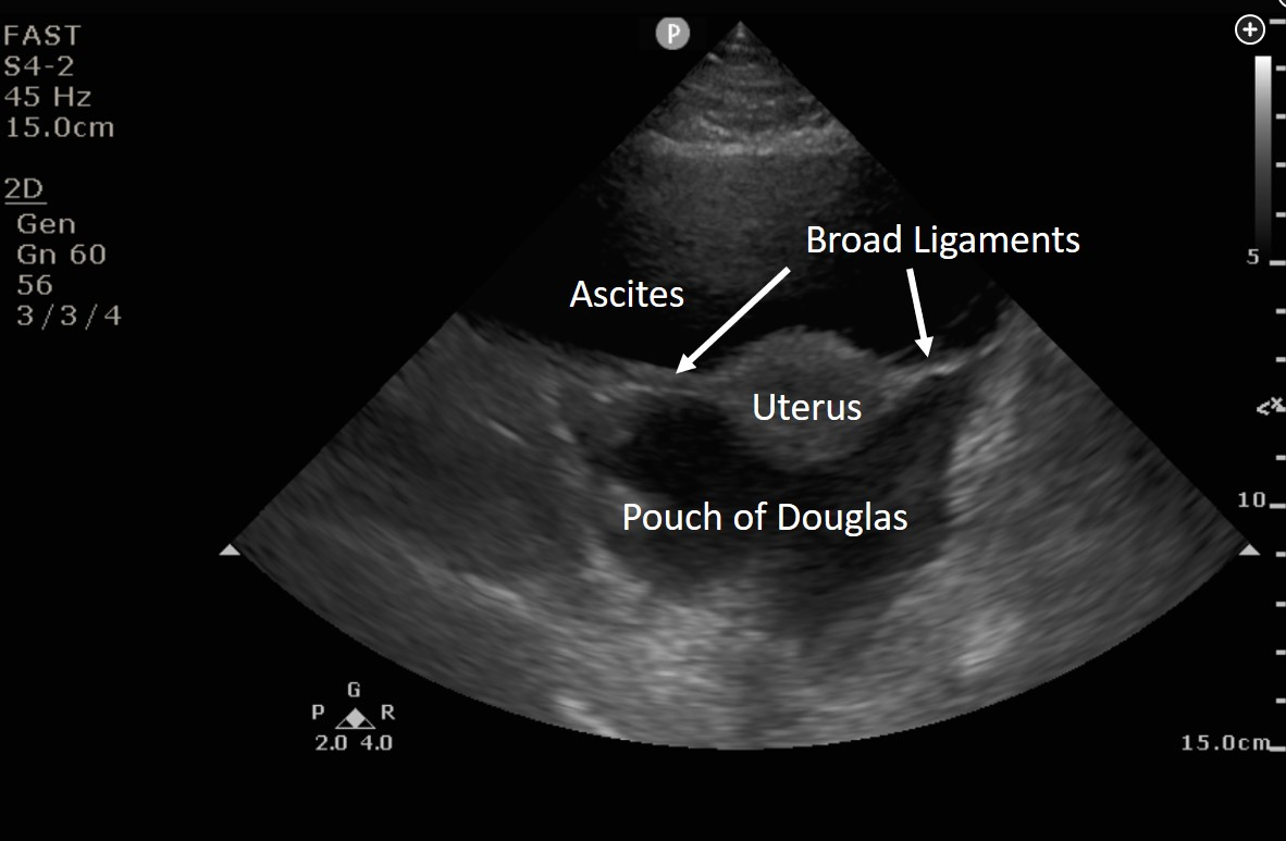
Interpretation of the FAST exam relies on knowledge of abdominal anatomy. Figure 2 is a transverse view just above the symphysis pubis showing the uterus suspended in anechoic fluid. The star is located posterior to the uterus in the pouch of Douglas, also called the rectouterine pouch (choice B is correct). The urinary bladder is located anterior to the uterus, not posterior (choice A is incorrect). The sigmoid colon should not present as an anechoic structure (choice C is incorrect). The bifurcation of the inferior vena cava should be much smaller than the area shown and would not be seen this close to the uterus (choice D is incorrect). Whenever there is doubt about whether an anechoic structure is vascular, color Doppler imaging can be helpful.123
Links to this note
Footnotes
-
Hamada SR, Delhaye N, Kerever S, et al. Integrating eFAST in the initial management of stable trauma patients: the end of plain film radiography. Ann Intensive Care. 2016;6(1):62. PubMed ↩
-
Von Kuenssberg Jehle D, Stiller G, Wagner D. Sensitivity in detecting free intraperitoneal fluid with the pelvic views of the FAST exam. Am J Emerg Med. 2003;21(6):476-478. PubMed ↩