middle mediastinum mass
- related: lung mass and cancer
- tags: #literature #pulmonology

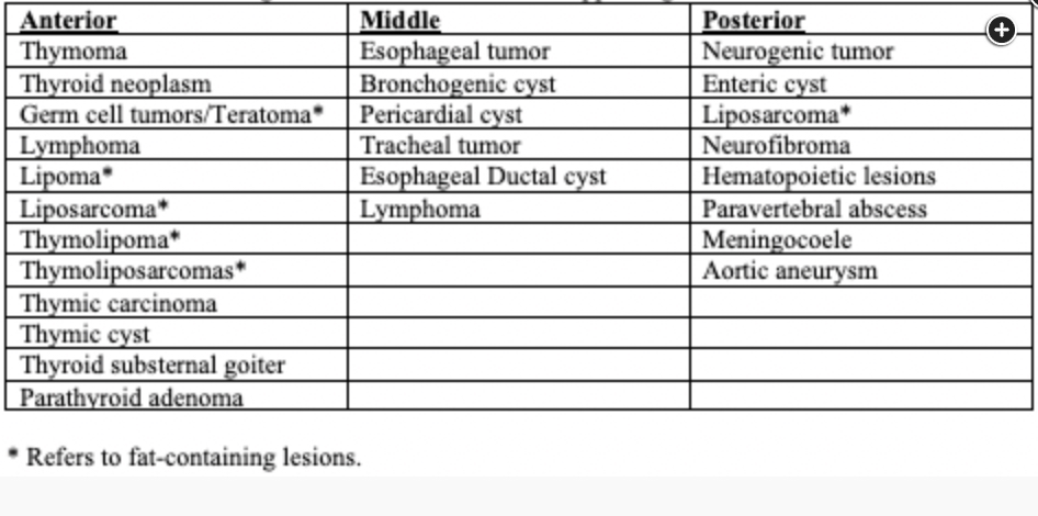
The most common middle mediastinal masses belong in one of three “A” categories—adenopathy, aneurysm, and abnormalities of development. Many other less common causes have been reported. Adenopathy may be present due to neoplasm (including lymphoma or carcinoma), infections (most commonly fungal or mycobacterial), and sarcoidosis. Abnormalities of development include bronchogenic cysts, esophageal duplication cysts, and pericardial cysts. Bronchogenic cysts are often found adjacent to the lower trachea or proximal mainstem bronchi while pericardial cysts are most commonly seen in the right cardiophrenic angle. Cysts are typically the attenuation of water on CT imaging though they can have a higher density after infection or hemorrhage.
It is important to recognize when a mediastinal mass is hypervascular (ie, enhancing on contrast imaging). This helps to narrow the differential diagnosis and appropriately plan when biopsy is needed. Hypervascular masses that involve the middle mediastinum include angiofollicular lymph node hyperplasia (Castleman’s disease), vascular malformations, metastatic disease, and paragangliomas. Hypervascular lymph node metastases usually originate from hypervascular tumors (eg, renal cell carcinoma, melanoma, thyroid carcinomas, and neuroendocrine tumors). Hypervascular masses in other mediastinal compartments include the previously mentioned plus ectopic parathyroid adenomas, thymic carcinoids (anterior mediastinum), and neurogenic tumors (posterior mediastinum).1