paragangliomas is a type of middle mediastinum mass
- related: lung mass and cancer
- tags: #literature #pulmonology
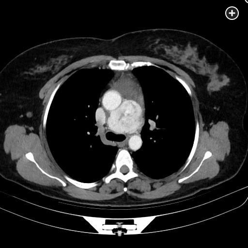
The mediastinal windows of the chest CT scan with contrast show an enhancing mass in the middle mediastinum. Paragangliomas can diffusely enhance on CT imaging while none of the other conditions listed do so (hodgkin lymphoma, bronchogenic cyst).
Paragangliomas arise from chromaffin cells in the adrenal glands and from extra-adrenal neuroendocrine tissues. In the adrenal glands, they are known as “pheochromocytomas.” Mediastinal paraganglia are present in the aortic body and in the aortopulmonary window in the middle mediastinum (branchiomeric paraganglia including the aortopulmonary paraganglia), and along the sympathetic trunk in the posterior mediastinum (aortosympathetic paraganglia). Paragangliomas arising in the middle mediastinum are more likely to be asymptomatic and occur in those ≥40 years old. Their presentation is delayed until they develop chest pain or shortness of breath due to compression of nearby structures. They are typically located adjacent to the great vessels. They homogeneously and avidly enhance on CT imaging when small and may be more heterogeneous when larger. Those in the posterior mediastinum more often present with symptoms and testing similar to an adrenal pheochromocytoma. A mutation in the subunit B, C, or D mitochronidrial succinate dehydrogenase gene of those with a paraganglioma is associated with earlier presentation and a greater risk of malignant transformation and metastatic disease. Rare presentations include Carney’s triad, which includes pulmonary chondroma and gastrointestinal stromal tumor, and the Von Hippel-Lindau syndrome. Treatment is surgical resection with curative intent. Invasion of nearby structures, such as the great vessels, may limit the ability to completely resect the tumor.1