terminologies and ranges in CPET testing
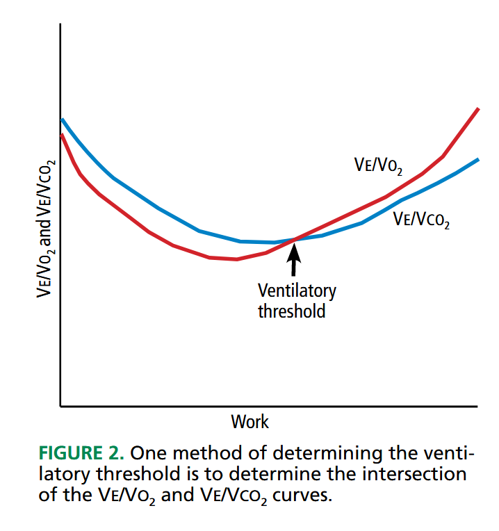
- Ve: ventilatory volume or minute ventilation. Volume of air expired1
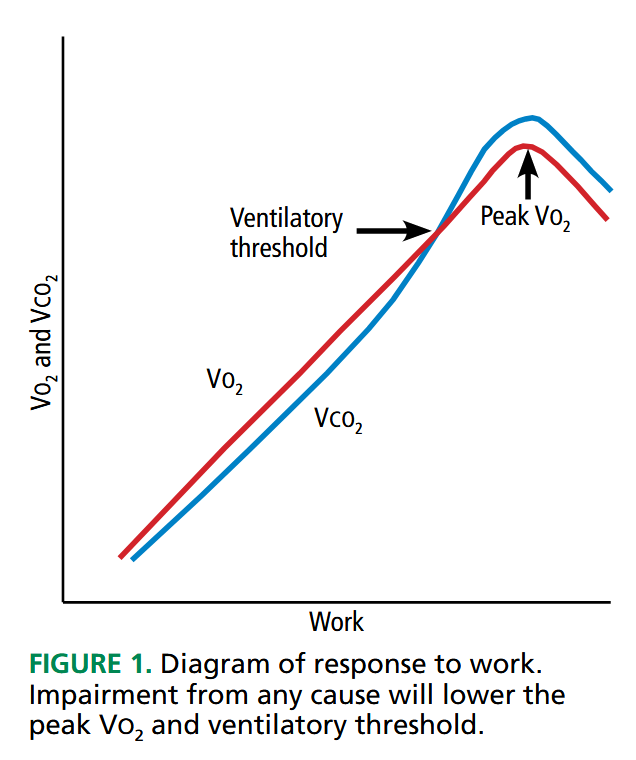
- VO2: volume of oxygen obtained
- peak VO2: highest oxygen uptake. Max achievable uptake
- marker of aerobic capacity2
- generally ≥ 85% predicted. The higher the better
- ≤ 84% is deemed abnormal3
- < 20 mL/kg/min in elderly
- < 14 mL/kg/min = poor prognosis in heart failure
- ≤ 10 if on beta blockers
- VCO2 volume of carbon dioxide expired. Typically increases with VO2 increases until ventilatory threshold is reached then increases more rapidly.4
- VO2θ anaerobic threshold: point of anaerobic metabolism
- VO2 usually at 40-60% peak VO2
- high value: athletic training
- low value: deconditioning
- VEθ ventilatory threshold: point at which buffer system is not enough to keep up with CO2 production, blood pH falls. ETCO2 increases.5
- MVV: maximum voluntary ventilations
- peak Ve/MVV: ventilatory reserve
- 15-20%
- reduced in athletes
- otherwise reduced in pulmonary limitation
- high in submaximal effort
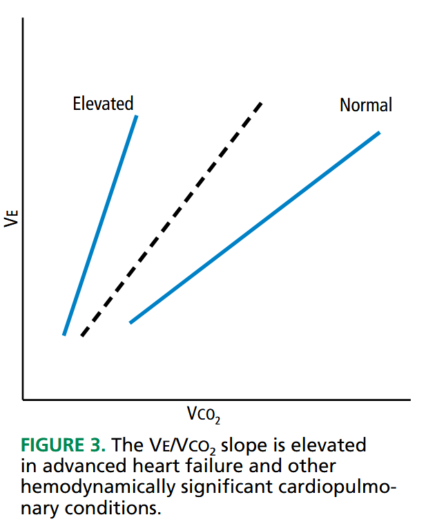
- Ve/VCO2 slope: ventilatory efficiency, marker of V/Q mismatch. Increased slope = worse disease
- normal 25-30
- ≥ 34 = significant cardiopulmonary disease
- VO2/work slope: oxygen uptake per unit of work
- normal 10±1.5 mL/min/watt
- high slope: increased anaerobic demand, high O2 cost (obesity, hyperthyroidism)
- low slope: increased anaerobic work (heart failure, CAD)
- VO2/kg: low = maybe obesity related6


Links to this note


